eVidyarthi
Menu
  • School
    • Close
    • CBSE English Medium
    • CBSE Hindi Medium
    • UP Board
    • Bihar Board
    • Maharashtra Board
    • MP Board
    • Close
  • Sarkari Exam Preparation
    • Close
    • Notes For Competitive Exams
    • MCQs for Competitive Exams
    • All Govt Exams Preparation
    • NCERT Syllabus for Competitive Exam
    • Close
  • Study Abroad
    • Close
    • Study in Australia
    • Study in Canada
    • Study in UK
    • Study in Germany
    • Study in USA
    • Close
Biology Class 11 Maharashtra Board | Menu
  • MCQ Biology Class 11 Maharashtra Board
  • Question Answers Biology Class 11 Maharashtra Board
  • Notes Biology Class 11 Maharashtra Board
  • Important Questions Biology Class 11 Maharashtra Board
  • Book Biology Class 11 Maharashtra Board
  • Biology Class 11

Within Chapter Questions Class 11 Chapter 16 Biology महाराष्ट्र Board

Skeleton and Movement

Can you recall? Page No. 193

1. Which are different types of muscular tissues ?

Answer: The different types of muscular tissues are smooth muscles, cardiac muscles, and striated (skeletal) muscles.

2. Name the type of muscles which bring about running and speaking.

Answer: Striated (skeletal) muscles bring about running and speaking, as they control voluntary movements.

3. Name the muscles which do not contract as per of our will.

Answer: Smooth muscles and cardiac muscles do not contract as per our will, as they are involuntary.

4. Which types of muscles show rhythmic contractions?

Answer: Cardiac muscles show rhythmic contractions, controlling the heart’s beating.

5. Which type of muscle is present in the diaphragm of the respiratory system?

Answer: Striated (skeletal) muscle is present in the diaphragm, allowing voluntary control during breathing.

6. Name the part of human skeleton situated along the vertical axis.

Answer: The axial skeleton, including the skull, vertebral column, and thoracic cage, is situated along the vertical axis.


Think about it Page No. 193

1. Why do we shiver during winter ?

Answer: Shivering occurs in winter as skeletal muscles contract rapidly and involuntarily to generate heat through muscle activity, helping to maintain body temperature in cold conditions.

2. Why do muscles show spasm after rigourous contraction?

Answer: Muscles show spasms after rigorous contraction due to the accumulation of lactic acid from anaerobic metabolism, causing fatigue and temporary disruption in muscle relaxation, leading to involuntary contractions.

3. Did you ever feel tickling in muscles?

Answer: The document does not directly address the sensation of tickling in muscles, but this feeling may occur due to nerve irritation or minor muscle contractions, often linked to fatigue or stimulation of sensory nerves in the muscle tissue.


Can you tell? Page No. 194

1. Why are movement and locomotion necessary among animals?

Answer: Movement and locomotion are necessary among animals to search for food, find shelter, locate mates, reach breeding grounds, and escape from predators, ensuring survival and reproduction.

2. Differentiate between :

Answer:

a. Flexor and Extensor Muscles

  • Flexor Muscles: These muscles contract to bend or flex a joint, reducing the angle between bones, e.g., biceps flex the elbow.
  • Extensor Muscles: These muscles contract to straighten or extend a joint, increasing the angle between bones, e.g., triceps extend the elbow.

b. Pronator and Supinator

  • Pronator Muscles: These muscles rotate the forearm to turn the palm downward, e.g., pronator teres.
  • Supinator Muscles: These muscles rotate the forearm to turn the palm upward, e.g., supinator muscle.

3. What are antagonistic muscles? Explain with example.

Answer: Antagonistic muscles are pairs of muscles that produce opposite actions at a joint, working together to control movement. For example, the biceps (flexor) and triceps (extensor) in the upper arm are antagonistic; the biceps contract to bend the elbow, while the triceps contract to straighten it, ensuring coordinated movement.


Can you tell? Page No. 197

1. Why are muscle rich in creatine phosphate?

Answer: Muscles are rich in creatine phosphate because it serves as a rapid energy reserve, quickly transferring phosphate to ADP to regenerate ATP, which is essential for muscle contraction during short bursts of intense activity.

2. What do you understand by muscle twitch?

Answer: A muscle twitch is a single, brief contraction and relaxation of a muscle fiber initiated by a single electrical stimulus, consisting of a latent period (no contraction), a contraction period, and a relaxation period.

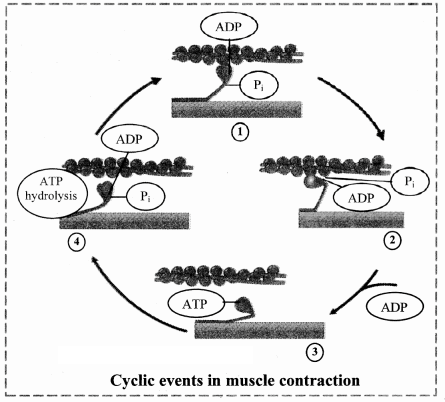
3. Explain mechanism of muscle contraction and relaxation.

Answer:

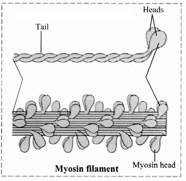
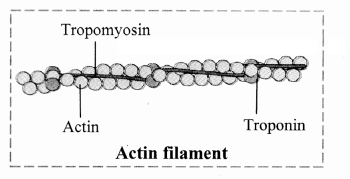
  • Contraction: A nerve impulse triggers the release of calcium ions from the sarcoplasmic reticulum, which bind to troponin, shifting tropomyosin to expose actin’s binding sites. Myosin heads, energized by ATP hydrolysis, bind to actin, forming cross-bridges, and tilt to pull actin filaments inward, shortening the sarcomere.
  • Relaxation: When stimulation stops, calcium is pumped back into the sarcoplasmic reticulum using ATP, allowing the troponin-tropomyosin complex to cover actin’s binding sites again. Myosin detaches from actin (using ATP), and actin filaments slide back, relaxing the muscle.

4. Explain the chemical changes taking place in muscle contraction.

Answer: During muscle contraction, ATP is hydrolyzed by myosin heads to ADP and inorganic phosphate, releasing energy for cross-bridge formation and the power stroke, pulling actin filaments. Calcium ions bind to troponin, triggering a conformational change in tropomyosin to expose actin’s binding sites. In relaxation, ATP is used to pump calcium back into the sarcoplasmic reticulum and detach myosin from actin, restoring the resting state.


Do you remember? Page No. 198

1. What are the components of our skeletal system ?

Answer: The skeletal system consists of bones, cartilage, and exoskeletal structures like nails, horns, hooves, scales, and hair, with bones and cartilage forming the major endoskeletal components.

2. What type of bones are present in our body?

Answer: The human body contains long bones (e.g., femur), short bones (e.g., carpals), flat bones (e.g., skull), irregular bones (e.g., vertebrae), and sesamoid bones (e.g., patella).

3. How do bones help us in various ways ?

Answer: Bones provide structural support and shape to the body, protect delicate organs (e.g., skull protects the brain), facilitate movement through joints, serve as attachment sites for muscles, store calcium, and act as sites for hematopoiesis (blood cell formation).


Identify and label Page No. 199

Identify the different bones.
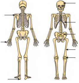
Answer:


Use your brain power Page No. 199

Why are long bones slightly bent and not straight ?

Answer: Long bones are slightly bent and not straight to distribute weight and stress more evenly across their structure, enhancing their strength and flexibility to withstand mechanical forces during movement and weight-bearing activities.


Can you tell? Page No. 201

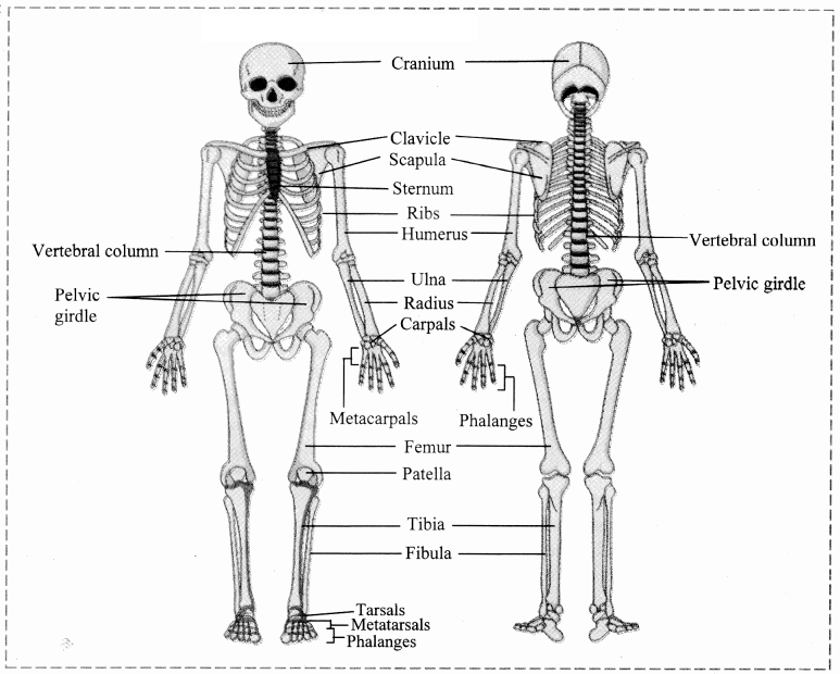
1. Give schematic plan of human skeleton.

Answer: The human skeleton consists of 206 bones, divided into two main divisions:

  • Axial Skeleton (80 bones): Includes the skull (22 bones: 8 cranium, 14 facial), hyoid bone (1), ear ossicles (6), vertebral column (26: 7 cervical, 12 thoracic, 5 lumbar, 1 sacrum, 1 coccyx), and thoracic cage (25: 1 sternum, 24 ribs).
  • Appendicular Skeleton (126 bones): Comprises the pectoral girdle (4: 2 clavicles, 2 scapulae), upper limbs (60: 2 humerus, 2 radius, 2 ulna, 16 carpals, 10 metacarpals, 28 phalanges), pelvic girdle (2: 2 coxal bones), and lower limbs (60: 2 femur, 2 patella, 2 tibia, 2 fibula, 14 tarsals, 10 metatarsals, 28 phalanges).

2. Enlist the bones of cranium.

Answer: The bones of the cranium are:

  • Frontal bone (unpaired)
  • Parietal bones (paired)
  • Temporal bones (paired)
  • Occipital bone (unpaired)
  • Sphenoid bone (unpaired)
  • Ethmoid bone (unpaired)

3. Write a note on structure and function of skull.

Answer:

  • Structure: The skull comprises 22 bones, divided into the cranium (8 bones) and facial bones (14 bones). The cranium includes the frontal, parietal, temporal, occipital, sphenoid, and ethmoid bones, forming a protective brain box with sutures (immovable joints) like coronal, sagittal, lambdoidal, and lateral/squamous. Facial bones, such as the maxilla, mandible, nasals, and zygomatic, shape the face, with the mandible being the only movable bone. The skull also includes features like the foramen magnum (for spinal cord passage) and sella turcica (for pituitary gland).
  • Function: The skull protects the brain, supports sensory organs (e.g., eyes, ears), provides attachment sites for muscles of the head and neck, and forms the framework for the face, contributing to identity and functions like chewing and speech. It also houses sinuses to lighten the skull and aid voice resonance.

Can you Tell? Page No. 202

Why skull is important for us? Enlist few reasons.

Answer: The skull is crucial for several reasons:

  • It protects the brain from injury, ensuring the safety of this vital organ.
  • It provides structural support and shape to the face, contributing to individual identity.
  • It houses sensory organs (e.g., eyes, ears) and supports functions like vision and hearing.
  • It serves as an attachment site for muscles involved in head movement, chewing, and speech.
  • It contains sinuses that lighten the skull and aid in voice resonance.

Try this Page No. 202

Feel your spine (vertebral column). Is it straight or curved?

Answer: The vertebral column is not straight; it has four curvatures: cervical and lumbar curves (secondary, convex) and thoracic and sacral curves (primary, concave). These curvatures help maintain balance in an upright position, absorb shocks during walking, and protect vertebrae from fracture.


Can you tell? Page No. 204

1. Explain the structure of a typical vertebra.

Answer: A typical vertebra consists of a prominent central body called the centrum, which is flat in the anteroposterior aspect (amphiplatyan). Posterior to the centrum, two short, thick processes unite to form the neural arch, creating a vertebral foramen that surrounds the spinal cord. The neural arch extends into a spinous process (neural spine) for muscle attachment. Transverse processes project laterally from the neural arch for muscle attachment. Superior and inferior zygapophyses (articular processes) on either side allow articulation with adjacent vertebrae, and intervertebral foramina at their junctions permit passage of spinal nerves.

2. How will you identify a thoracic vertebra?

Answer: A thoracic vertebra can be identified by its heart-shaped centrum and well-developed processes. It has facets on the transverse processes (except for vertebrae 11 and 12) for articulation with ribs, and the centrum has demifacets for rib attachment. These features, along with its location in the chest region, distinguish it from other vertebrae.

3. Write a note on curvatures of vertebral column and mention their importance.

Answer: The vertebral column has four curvatures: cervical and lumbar (secondary, convex, developing after birth) and thoracic and sacral (primary, concave, present at birth). These curvatures maintain balance in an upright posture, distribute mechanical stress evenly, and absorb shocks during walking or other activities. They also protect the vertebrae from fracture and enhance the spine’s flexibility and resilience.


Can you recall? Page No. 206

How does humerus form ball and socket joint? Where is it located ?

Answer: The humerus forms a ball and socket joint through its hemispherical head at the proximal end, which fits into the glenoid cavity of the scapula, a concave socket in the pectoral girdle. This structure allows multiaxial movements, including flexion, extension, abduction, adduction, and rotation. The joint is located at the shoulder, connecting the upper arm to the axial skeleton.


Can you tell? Page No. 208

1. Differentiate between skeleton of palm and foot.

Answer:

Skeleton of palmSkeleton of foot
a.It consists of metacarpals and phalangesIt consists of metatarsals and phalanges
b.Saddle joints and condyloid joints are in the palm.Condyloid or saddle joints are not present the foot.

2. Explain the longest bone in human body.

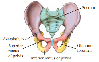
Answer: The femur, located in the thigh, is the longest bone in the human body. It has a head connected to the shaft by a short neck, forming a ball and socket joint with the acetabulum of the coxal bone at the hip. The shaft’s lower third forms a triangular popliteal surface, and the distal end has two condyles that articulate with the tibia and fibula, supporting weight-bearing and movement.


Do you remember? Page No. 208

1. What are joints? What are their types?

Answer: Joints, also known as articulations or arthroses, are points where two or more bones meet, enabling movement and flexibility. They are classified based on the degree of movement into three types:

  • Synarthroses (Fibrous Joints): Immovable, held by fibrous connective tissue, e.g., sutures (skull), syndesmoses (tibiofibular ligament), gomphoses (teeth in jaw).
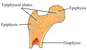
  • Amphiarthroses (Cartilaginous Joints): Slightly movable, connected by hyaline or fibrocartilage, e.g., synchondroses (epiphyseal plate), symphysis (intervertebral discs, pubic symphysis).
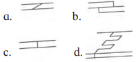
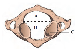
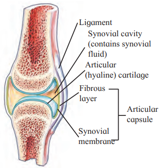
  • Diarthroses (Synovial Joints): Freely movable, with a synovial cavity, hyaline cartilage, and synovial fluid, e.g., pivot (atlas-axis), ball and socket (shoulder), hinge (elbow), condyloid (metacarpophalangeal), saddle (thumb), gliding (intercarpal).

2. What types of joint is present at knee?

Answer: The knee joint is a hinge joint, a type of synovial joint that allows monoaxial movement, primarily flexion and extension, between the femur and tibia.


Use your brain power Page No. 210

Why are warming up rounds essential before regular exercise?

Answer: Warming up rounds are essential before regular exercise because they increase blood flow to muscles, raise muscle temperature, and reduce the viscosity of synovial fluid in joints, thereby enhancing flexibility, improving performance, and preventing injuries.


Can you tell? Page No. 211

1. Human beings can hold an object in a better manner than monkeys. Why?

Answer: Human beings can hold objects better than monkeys due to the presence of a saddle joint at the carpometacarpal joint of the thumb, which allows for greater flexibility and precision in movements like opposition, abduction, and circumduction. This joint, characteristic of Homo sapiens, enables a stronger and more precise grip compared to monkeys, whose thumb joints are less specialized for such fine motor tasks. Additionally, humans have more developed fine motor control and dexterity due to advanced neural coordination.

3. What makes the synovial joint freely moveable?

Answer: Synovial joints are freely movable due to the presence of a synovial cavity filled with synovial fluid, which lubricates the joint and reduces friction. The articulating surfaces are covered with hyaline cartilage, which minimizes wear and absorbs shocks, while the synovial membrane and ligaments (including the fibrous capsule) provide stability without restricting movement. These features collectively allow a wide range of motions, such as flexion, extension, rotation, and circumduction, depending on the joint type.

Important Questions Class 11 Chapter 16 Biology महाराष्ट्र Board

Skeleton and Movement

Short Questions

1. What is the functional unit of striated muscle?

Answer: The sarcomere is the functional unit of striated muscle.

2. Which bones are involved in an ankle fracture?

Answer: Tarsal bones are involved in an ankle fracture.

3. What causes muscle fatigue?

Answer: Accumulation of lactic acid causes muscle fatigue.

4. Which muscle pair is not antagonistic?

Answer: Sphincters and supinators are not an antagonistic pair.

5. How does soaking a sprained foot in salty water reduce swelling?

Answer: Osmosis draws water out of the swollen tissue, reducing swelling.

6. What is the role of calcium in muscle contraction?

Answer: Calcium binds to troponin, exposing actin’s binding sites for myosin.

7. Which disorder is caused by hyper-secretion of parathormone?

Answer: Osteoporosis is caused by hyper-secretion of parathormone.

8. What type of contraction occurs in neck muscles while reading?

Answer: Isometric contraction occurs in neck muscles while reading.

9. Which joint moves during bicep training with dumbbells?

Answer: The elbow joint moves during bicep training with dumbbells.

10. Why is a splint essential for a fractured leg?

Answer: A splint immobilizes the fracture, preventing further damage and pain.

11. Why is a sprain more painful than a fracture?

Answer: Sprains involve nerve-rich ligaments, causing intense pain and inflammation.

12. Why do red muscle fibers work longer than white fibers?

Answer: Red fibers use aerobic respiration, resisting fatigue longer than white fibers.

13. What is the total number of bones in the human skeleton?

Answer: The human skeleton has 206 bones.

14. What type of joint is the elbow?

Answer: The elbow is a hinge joint, allowing flexion and extension.

15. What is rigor mortis?

Answer: Rigor mortis is post-death muscle stiffening due to ATP depletion.

Long Questions

1. How does the sarcomere’s structure facilitate muscle contraction?

Answer: The sarcomere contains actin and myosin filaments arranged for sliding. Calcium triggers tropomyosin movement, exposing actin’s binding sites. Myosin heads pull actin, shortening the sarcomere via the sliding filament theory.

2. Why did Ragini, a 50-year-old, suffer hairline cracks in her feet?

Answer: Ragini likely has osteoporosis due to postmenopausal estrogen decline. Low calcium and vitamin D intake weakened her bones. She should take calcium supplements and exercise to prevent further fractures.

3. How do actin and myosin structures contribute to muscle contraction?

Answer: Actin’s tropomyosin and troponin regulate myosin binding when calcium is present. Myosin’s heads, with ATPase activity, form cross-bridges with actin. These bridges pull actin, causing sarcomere shortening and contraction.

4. Why are the atlas and axis vertebrae structured differently?

Answer: The atlas is ring-like, supporting the skull and enabling nodding. The axis’s odontoid process forms a pivot joint for rotation. Their structures suit their roles in head movement and spinal cord protection.

5. Why is a wooden splint used for a fractured leg in emergencies?

Answer: A splint stabilizes the fractured leg, preventing bone movement. This reduces pain and further tissue damage during transport. It ensures safe handling until medical treatment is provided.

6. Why are sprains often more painful than fractures?

Answer: Sprains damage ligaments, which are richly innervated, causing severe pain. Fractures may be less painful if the break is clean. Swelling and inflammation in sprains further intensify discomfort.

7. How do red and white muscle fibers differ in function?

Answer: Red fibers, rich in myoglobin and mitochondria, support prolonged aerobic activity. White fibers, relying on anaerobic glycolysis, fatigue quickly due to lactic acid buildup. This suits red fibers for endurance and white for short bursts.

8. What is the role of calcium in muscle contraction and relaxation?

Answer: Calcium binds to troponin, exposing actin’s binding sites for contraction. During relaxation, calcium is pumped back into the sarcoplasmic reticulum. This restores tropomyosin’s position, preventing myosin-actin interaction.

9. How do synovial joints enable free movement?

Answer: Synovial joints have a synovial cavity filled with lubricating fluid. Hyaline cartilage reduces friction, and ligaments prevent dislocation. This structure allows multiaxial or monoaxial movements, like in the hip or elbow.

10. What is osteoporosis, and how can it be prevented?

Answer: Osteoporosis is a condition where bones become brittle due to excessive resorption. It can be prevented with a calcium-rich diet and weight-bearing exercises. Regular bone density screenings help monitor and manage risk.

Notes Class 11 Chapter 16 Biology महाराष्ट्र Board

Skeleton and Movement

Introduction

Organisms exhibit various movements, from protoplasmic streaming to locomotion like walking or running. Movements can be internal (e.g., peristalsis) or external (e.g., limb movement) and may be voluntary or involuntary. Locomotion involves the displacement of an organism’s entire body, driven by the need for food, shelter, mates, or escaping predators. This chapter explores the skeletal system, muscles, and their roles in movement and locomotion in humans.


16.1 Movements and Locomotion

Movements

Movements are classified as:

  • Internal: Occur within the body, e.g., peristalsis in the alimentary canal, heartbeat.
  • External: Involve body parts, e.g., limb movements, head rotation.
  • Voluntary: Controlled consciously, e.g., walking, writing.
  • Involuntary: Not under conscious control, e.g., heartbeat, peristalsis.

Types of Muscles and Their Roles:

  1. Smooth Muscles: Involuntary, control internal movements like peristalsis, blood vessel constriction/dilation.
  2. Cardiac Muscles: Involuntary, responsible for heart contraction and relaxation.
  3. Striated (Skeletal) Muscles: Voluntary, control movements of limbs, head, trunk, etc.

Examples of Movements:

  • Whorling Movement: Performed by flagella, e.g., sperm movement.
  • Peristaltic Movement: Wave-like contractions in the alimentary canal.
  • Eye Movement: Controlled by striated muscles for voluntary gaze shifts.

Locomotion

Locomotion is the movement of an organism from one place to another, driven by survival needs (e.g., finding food, escaping enemies). It involves coordinated action of bones, joints, and muscles.

Types of Locomotory Movements:

  1. Amoeboid Movement: Via pseudopodia, e.g., leucocytes.
  2. Ciliary Movement: Via cilia, e.g., ciliated epithelium in Paramecium aiding food passage.
  3. Whorling Movement: Via flagella, e.g., sperm.
  4. Muscular Movement: Via muscles, bones, and joints, e.g., walking, running.

Key Fact: All locomotions are movements, but not all movements result in locomotion.

Human Muscle Facts:

  • Approximately 640 muscles in the human body (634 paired, 6 unpaired).
  • Skeletal muscles are attached to bones via tendons (inelastic collagen fiber bands).

16.2 Location and Structure of Skeletal Muscles

Skeletal muscles are striated, voluntary muscles attached to bones, enabling movement. They are typically located on a bone proximal to the one they move (e.g., biceps/triceps in the upper arm move the forearm).

Structure

  • Origin: The end of the muscle attached to a stationary bone.
  • Insertion: The end attached to a movable bone.
  • Belly: The thick, central part of the muscle, fusiform in shape due to the maximum number of fibers in the middle.

Types of Skeletal Muscles (Based on Movement)

  1. Agonists (Prime Movers): Initiate movement, e.g., biceps for forearm flexion.
  2. Antagonists: Produce opposite action, e.g., triceps for forearm extension.
  3. Synergists: Assist prime movers, e.g., brachialis assists biceps.

Muscle Arrangement

  • Muscles work in antagonistic pairs, where one muscle’s contraction opposes the other’s action (e.g., biceps flex the elbow, triceps extend it).
  • One muscle in a pair is typically stronger (e.g., biceps stronger than triceps).
  • Muscles can only pull (contract), not push, due to their contractile nature.

16.3 Working of Skeletal Muscles

Skeletal muscles produce movement by contracting and pulling bones at joints. They work in antagonistic pairs to enable opposing actions (e.g., flexion and extension).

Antagonistic Muscle Pairs

  1. Flexor and Extensor:
    • Flexor: Bends a joint, e.g., biceps.
    • Extensor: Straightens a joint, e.g., triceps.
  2. Abductor and Adductor:
    • Abductor: Moves a part away from the body axis, e.g., deltoid (shoulder).
    • Adductor: Moves a part toward the body axis, e.g., latissimus dorsi.
  3. Pronator and Supinator:
    • Pronator: Turns the palm downward.
    • Supinator: Turns the palm upward.
  4. Levator and Depressor:
    • Levator: Raises a body part.
    • Depressor: Lowers a body part.
  5. Protractor and Retractor:
    • Protractor: Moves a part forward.
    • Retractor: Moves a part backward.
  6. Sphincters: Circular muscles controlling openings, e.g., in the anus or stomach.

16.4 Mechanism of Muscle Contraction

Sarcomere: The Contractile Unit

The sarcomere is the functional unit of striated muscle, containing:

  • Actin Filaments (thin): Anchored at Z-lines, with tropomyosin and troponin.
  • Myosin Filaments (thick): Centrally located, with heads forming cross-bridges.

Structure of Contractile Proteins

  1. Myosin Filament:
    • Composed of multiple meromyosin units.
    • Each unit has two heavy chains (forming a tail and head) and four light chains.
    • Myosin head: Has ATPase activity, splits ATP for energy, and forms cross-bridges with actin.
    • Tails point toward the sarcomere center, heads outward.
  2. Actin Filament:
    • F-actin: Double-stranded polymer of G-actin molecules (each with ADP).
    • Tropomyosin: Covers myosin-binding sites in the resting state.
    • Troponin: Binds calcium, tropomyosin, and actin, initiating contraction when calcium is present.

Sliding Filament Theory

Proposed by H.E. Huxley and A.F. Huxley, this theory explains muscle contraction:

  1. Action Potential: A nerve impulse triggers calcium release from the sarcoplasmic reticulum.
  2. Calcium Binding: Calcium binds to troponin, shifting tropomyosin to expose actin’s binding sites.
  3. Cross-Bridge Formation: Myosin heads, energized by ATP hydrolysis, bind to actin, forming an actomyosin complex.
  4. Power Stroke: Myosin heads tilt, pulling actin filaments inward, shortening the sarcomere.
  5. Detachment: ATP binds to myosin, releasing it from actin, and is hydrolyzed for the next cycle.

This sliding of actin over myosin shortens the muscle fiber, causing contraction.


16.5 Physiology of Muscle Relaxation

  • In the relaxed state, tropomyosin and troponin cover actin’s binding sites, preventing myosin interaction.
  • Relaxation Process:
    1. Stimulation ceases, stopping calcium release.
    2. Calcium is actively pumped back into the sarcoplasmic reticulum (using ATP).
    3. Troponin-tropomyosin complex reforms, covering actin’s binding sites.
    4. Myosin detaches from actin (using ATP), and actin filaments slide back, relaxing the muscle.
  • Relaxation is an active process requiring ATP, similar to contraction.

16.6 Properties of Muscles on Electrical Stimulation

  1. Single Muscle Twitch: A brief contraction from one stimulus, with three stages:
    • Latent Period: No contraction.
    • Contraction Period: Muscle shortens.
    • Relaxation Period: Muscle returns to original length.
  2. Summation: Repeated stimuli before twitch completion increase contraction strength (staircase phenomenon).
  3. Tetanus: Rapid, frequent stimuli prevent relaxation, maintaining contraction.
  4. Refractory Period: A brief period (~0.02 seconds) after a stimulus where the muscle cannot respond to another.
  5. Threshold Stimulus: Minimum stimulus strength required for contraction.
  6. All or None Principle: A muscle fiber contracts fully or not at all, depending on whether the stimulus reaches the threshold.
  7. Oxygen Debt: During strenuous exercise, muscles contract anaerobically, accumulating lactic acid. Post-exercise, extra oxygen (oxygen debt) oxidizes lactic acid and restores ATP/creatine phosphate.

Rigor Mortis: Post-death muscle stiffening due to ATP depletion, preventing myosin detachment from actin. It helps estimate time of death.


16.7 Skeletal System

The skeletal system provides structural support, protects organs, enables movement, stores calcium, and supports hematopoiesis. It consists of bones and cartilage, forming the endoskeleton (internal) and exoskeleton (external, e.g., nails, hair).

Human Skeleton

  • Total Bones: 206 in adults, divided into:
    • Axial Skeleton: 80 bones (along the body’s longitudinal axis).
    • Appendicular Skeleton: 126 bones (limbs and girdles).
  • Cartilage: Pliable, found in joints and certain skeletal parts (e.g., nose, ear).
  • Bones: Rigid, with a hard matrix, providing shape and support.

Comparison with Simple Machines

Bones, muscles, and joints function like a lever:

  • Joint: Acts as the fulcrum.
  • Muscle: Provides the effort (force).
  • Bone: Acts as the resistance (load).
  • Examples:
    • Class I Lever: Head movement at the atlanto-occipital joint (fulcrum: joint, effort: back muscles, resistance: head).
    • Class II Lever: Standing on toes (fulcrum: toe, effort: calf muscles, resistance: body).
    • Class III Lever: Forearm flexion at the elbow (fulcrum: elbow, effort: biceps, resistance: radius/ulna).

16.8 Axial Skeleton (80 Bones)

Skull (22 Bones)

  • Cranium (8 Bones):
    • Frontal Bone: Forehead, roof of orbits (unpaired).
    • Parietal Bones: Roof and sides of cranium (paired).
    • Temporal Bones: Lateral, above ears, with zygomatic process and mandibular fossa (paired).
    • Occipital Bone: Back and base, with foramen magnum and occipital condyles (unpaired).
    • Sphenoid Bone: Butterfly-shaped, holds cranial bones together, with sella turcica for pituitary (unpaired).
    • Ethmoid Bone: Spongy, supports nasal cavity, part of nasal septum (unpaired).
  • Facial Bones (14 Bones):
    • Nasals: Bridge of nose (paired).
    • Maxillae: Upper jaw, house upper teeth (paired).
    • Palatines: Roof of buccal cavity (paired).
    • Zygomatic Bones: Cheekbones, form zygomatic arch (paired).
    • Lacrimal Bones: Medial orbit wall, house lacrimal sacs (paired).
    • Inferior Nasal Conchae: Lateral nasal cavity wall (paired).
    • Vomer: Inferior nasal septum (unpaired).
    • Mandible: Lower jaw, only movable skull bone (unpaired).
  • Sutures: Immovable joints in the skull:
    • Coronal: Frontal to parietals.
    • Sagittal: Between parietals.
    • Lambdoidal: Parietals to occipital.
    • Lateral/Squamous: Parietal to temporal.
  • Fontanelles: Soft spots in newborn skulls, ossify by age two, allowing flexibility during birth and brain growth.

Hyoid Bone (1 Bone)

  • U-shaped, suspended from temporal bones, supports tongue and neck muscles, does not articulate with other bones.

Ear Ossicles (6 Bones)

  • Malleus, Incus, Stapes: Three tiny bones in each middle ear, transmit sound vibrations.

Vertebral Column (26 Bones in Adults)

  • Childhood: 33 vertebrae; in adults, 5 sacral fuse into sacrum, 4 coccygeal into coccyx.
  • Types of Vertebrae:
    • Cervical (7): Neck, includes atlas (1st) and axis (2nd).
    • Thoracic (12): Chest, articulate with ribs.
    • Lumbar (5): Abdominal, robust for support.
    • Sacral (5, fused): Form sacrum, in pelvic cavity.
    • Coccygeal (4, fused): Form coccyx, vestigial tailbone.
  • Curvatures:
    • Primary: Thoracic and sacral (concave, present at birth).
    • Secondary: Cervical and lumbar (convex, develop post-birth).
    • Functions: Balance, shock absorption, protect vertebrae.
  • Typical Vertebra Structure:
    • Centrum: Central body, amphiplatyan (flat surfaces).
    • Neural Arch: Forms vertebral foramen (houses spinal cord).
    • Spinous Process: Posterior projection for muscle attachment.
    • Transverse Processes: Lateral projections for muscles.
    • Zygapophyses: Superior/inferior articular processes for vertebra articulation.
    • Intervertebral Foramina: Allow spinal nerve passage.

Special Vertebrae:

  • Atlas: Ring-like, no centrum, forms “Yes joint” with occipital condyles.
  • Axis: Has odontoid process, forms “No joint” (pivot) with atlas.

Thoracic Cage (25 Bones)

  • Sternum (1):
    • Parts: Manubrium, body, xiphoid process.
    • Manubrium: Attaches to clavicles and first two rib pairs.
    • Body: Attaches to ribs via costal cartilages.
    • Xiphoid: Cartilaginous, ossifies in adults, attaches diaphragm.
  • Ribs (24, 12 pairs):
    • True Ribs (1-7): Directly attach to sternum via costal cartilages.
    • False Ribs (8-10): Attach to sternum indirectly via rib 7.
    • Floating Ribs (11-12): No sternal attachment.
    • Structure: Head (articulates with vertebrae), tubercle (attaches to transverse processes), costal groove.
    • Intercostal Spaces: House intercostal muscles.

16.9 Appendicular Skeleton (126 Bones)

Pectoral Girdle (4 Bones)

  • Clavicle (2): S-shaped, connects sternum to scapula, stabilizes shoulder.
  • Scapula (2): Flat, triangular, with glenoid cavity (for humerus), coracoid, and acromion processes for muscle attachment.

Upper Limbs (60 Bones)

  • Humerus (2): Upper arm, with head (forms shoulder joint), tubercles, bicipital groove, trochlea (articulates with ulna).
  • Radius (2): Lateral forearm, with disc-like head and styloid process.
  • Ulna (2): Medial forearm, with olecranon process (elbow joint) and radial notch.
  • Carpals (16): Wrist, 8 per hand, arranged in two rows.
  • Metacarpals (10): Palm, 5 per hand, form knuckles.
  • Phalanges (28): Fingers, 14 per hand (3 per finger, 2 per thumb).

Pelvic Girdle (2 Bones)

  • Coxal Bones (2): Each has ilium, ischium, pubis, fused at the acetabulum (hip joint).
  • Pubic Symphysis: Cartilaginous joint joining pubis bones.
  • Obturator Foramen: Space formed by pubis and ischium.

Lower Limbs (60 Bones)

  • Femur (2): Thigh, longest bone, with head (hip joint), neck, trochanters, condyles.
  • Patella (2): Knee cap, sesamoid bone.
  • Tibia (2): Medial shank, stronger, articulates with femur and talus.
  • Fibula (2): Lateral shank, slender, does not articulate with femur.
  • Tarsals (14): Ankle, 7 per foot (e.g., calcaneum, talus).
  • Metatarsals (10): Sole, 5 per foot.
  • Phalanges (28): Toes, 14 per foot (3 per toe, 2 for big toe).

16.10 Types of Joints

Joints (articulations) are points where bones meet, enabling movement. They are classified by movement degree:

1. Synarthroses (Fibrous, Immovable)

  • Held by fibrous connective tissue, no movement.
  • Types:
    • Sutures: Thin fibrous layer, e.g., skull sutures (coronal, sagittal, lambdoidal).
    • Syndesmoses: Fibrous tissue as sheets/bundles, e.g., tibiofibular ligament.
    • Gomphoses: Cone-shaped bone in a socket, e.g., teeth in jaw.

2. Amphiarthroses (Cartilaginous, Slightly Movable)

  • Held by hyaline or fibrocartilage, limited movement.
  • Types:
    • Synchondroses: Hyaline cartilage, e.g., epiphyseal plate, rib-sternum junction.
    • Symphysis: Fibrocartilage disc, e.g., intervertebral discs, pubic symphysis.

3. Diarthroses (Synovial, Freely Movable)

  • Feature a synovial cavity (filled with synovial fluid), hyaline cartilage on articulating surfaces, synovial membrane, and ligaments.
  • Synovial Fluid: Lubricates, absorbs shocks, nourishes cartilage, removes waste.
  • Types:
    • Pivot Joint: Rotation around one axis, e.g., atlas-axis (“No joint”).
    • Ball andзя- Socket Joint: Multiaxial movement, e.g., shoulder, hip.
    • Hinge Joint: Monoaxial (flexion/extension), e.g., elbow, knee.
    • Condyloid Joint: Biaxial (flexion/extension, abduction/adduction), e.g., metacarpophalangeal joint.
    • Saddle Joint: Biaxial, allows circumduction, e.g., thumb carpometacarpal joint.
    • Gliding Joint: Non-axial, flat surfaces, e.g., intercarpal joints.

16.11 Disorders Related to Muscles

  1. Muscular Dystrophy:
    • Genetic, progressive muscle wasting, affects voluntary muscles.
    • Types: Duchenne (boys, lower limbs), limb-girdle (adults, shoulders/hips).
    • No cure, weakens muscles over time.
  2. Myasthenia Gravis:
    • Autoimmune, weakens skeletal muscles by blocking acetylcholine receptors.
    • Symptoms: Ptosis, double vision, difficulty swallowing/speaking.
    • Causes progressive muscle weakness.

16.12 Disorders Related to Bones

  1. Arthritis: Joint inflammation, painful, can lead to disability.
    • Osteoarthritis: Cartilage degeneration, affects hands, knees, spine, due to aging, obesity.
    • Gouty Arthritis: Uric acid deposits in joints, causes inflammation, affects feet.
    • Rheumatoid Arthritis: Autoimmune, synovial membrane swells, forms pannus, erodes cartilage, stiffens joints.
  2. Osteoporosis:
    • Bones become porous and brittle, common in postmenopausal women.
    • Causes: Estrogen decline, low calcium/vitamin D, reduced bone formation.
    • Symptoms: Height loss, hunched back, bone pain, fractures.
    • Prevention: Calcium-rich diet, exercise, avoid smoking/alcohol.

Questions Answers Class 11 Chapter 16 Biology महाराष्ट्र Board

Skeleton and Movement

1. Choose the correct option

A. The functional unit of striated muscle is …………..
a. cross bridges b. myofibril
c. sarcomere d. z-band

Answer: c. sarcomere
(The sarcomere is the contractile unit of striated muscle, containing actin and myosin filaments responsible for muscle contraction.)

B. A person slips from the staircase and breaks his ankle bone. Which bones are involved?
a. Carpals b. Tarsal
c. Metacarpals d. Metatarsals

Answer: b. Tarsal
(The ankle consists of tarsal bones, which can be fractured in such an injury.)

C. Muscle fatigue is due to accumulation of ……..
a. pyruvic acid b. lactic acid
c. malic acid d. succinic acid

Answer: b. lactic acid
(Lactic acid accumulates during anaerobic glycolysis, causing muscle fatigue.)

D. Which one of the following is NOT antagonistic muscle pair?
a. Flexo-extensor
b. Adductor-abductor
c. Levator-depressor
d. Sphinetro-suprinater

Answer: d. Sphinctro-suprinater
(Sphincters and supinators do not form an antagonistic pair, unlike flexor-extensor, adductor-abductor, or levator-depressor.)

E. Swelling of sprained foot is reduced by soaking in hot water containing a large amount of common salt,
a. due to osmosis
b. due to plasmolysis
c. due to electrolysis
d. due to photolysis

Answer: a. due to osmosis
(The high salt concentration draws water out of the swollen tissue via osmosis, reducing swelling.)

F. Role of calcium in muscle contraction is ……….
a. to break the cross bridges as a cofactor in the hydrolysis of ATP
b. to bind with troponin, changing its shape so that the actin filament is exposed
c. to transmit the action potential across the neuromuscular junction.
d. to re-establish the polarisation of the plasma membrane following an action potential

Answer: b. to bind with troponin, changing its shape so that the actin filament is exposed
(Calcium binds to troponin, causing a conformational change that moves tropomyosin, exposing actin’s binding sites for myosin.)

G. Hyper-secretion of parathormone can cause which of the following disorders?
a. Gout b. Rheumatoid arthritis
c. Osteoporosis d. Gull’s disease

Answer: c. Osteoporosis
(Excess parathormone increases bone resorption, leading to porous and brittle bones, characteristic of osteoporosis.)

H. Select correct option between two nasal bones

Answer:


2. Answer the following questions

A. What kind of contraction occurs in your neck muscles while you are reading your class assignment?

Answer: Isometric contraction occurs in the neck muscles. These muscles contract to hold the head in a stable position without causing movement, maintaining posture during reading.

B. Observe the diagram and enlist importance of ‘A’, ‘B’ and ‘C’

Answer:

A – Posterior portion of vertebral foramen of atlas vertebrae; Importance – The spinal cord runs through this portion of vertebral foramen
B – Anterior portion of vertebral foramen of axis vertebrae; Importance – In this portion, the odontoid process of axis vertebrae forms ‘NO’ joint.
C – Inferior articular facet; Importance – It articulates with superior articular facet of axis and permits rotatory movement of head.

C. Raju intends to train biceps; while exercising using dumbbells, which joints should remain stationary and which should move?

Answer:

  • Stationary joint: The shoulder joint should remain relatively stationary to stabilize the upper arm.
  • Moving joint: The elbow joint should move, allowing flexion and extension as the biceps contract and relax during dumbbell exercises.

D. In a road accident, Moses fractured his leg. One of the passers by, tied a wodden plank to the fractured leg while Moseswas rushed to the hospital Was this essential? Why?

Answer: Yes, this was essential. Tying a wooden plank acts as a splint, immobilizing the fractured leg to prevent further movement of the broken bones. This reduces pain, prevents additional tissue damage, and minimizes the risk of complications like blood vessel or nerve injury during transport to the hospital.

E. Sprain is more painful than fracture. Why?

Answer: A sprain can be more painful than a fracture because it involves damage to ligaments, which are richly supplied with nerves, leading to intense pain. Fractures may sometimes cause less immediate pain if the break is clean and does not involve significant soft tissue damage. Additionally, sprains often result in swelling and inflammation, which further stimulate pain receptors.

F. Why a red muscle can work for a prolonged period whereas white muscle fibre suffers from fatigue after a shorter work? (Refer to chapter animal tissues.)

Answer: Red muscle fibers (slow-twitch) are rich in myoglobin and mitochondria, enabling efficient aerobic respiration and sustained energy production, allowing them to work for prolonged periods without fatigue. White muscle fibers (fast-twitch) rely on anaerobic glycolysis, leading to rapid energy production but quick accumulation of lactic acid, causing fatigue after shorter durations.


3. Answer the following questions in detail

A. How is the structure of sarcomere suitable for the contractility of the muscle? Explain its function according to sliding filament theory. (Refer to chapter animal tissues.)

Answer: The sarcomere, the functional unit of striated muscle, is highly organized to facilitate muscle contraction. Its structure includes:

  • Actin filaments (thin filaments) anchored at the Z-lines, forming the boundaries of the sarcomere.
  • Myosin filaments (thick filaments) located centrally, with heads that form cross-bridges with actin.
  • Tropomyosin and troponin on actin regulate the interaction with myosin.
    Sliding Filament Theory:

According to H.E. Huxley and A.F. Huxley’s sliding filament theory, muscle contraction occurs when actin and myosin filaments slide past each other, shortening the sarcomere without changing the length of the filaments themselves. The process involves:

  • Action Potential: A nerve impulse triggers the release of calcium ions from the sarcoplasmic reticulum into the sarcoplasm.
  • Calcium Binding: Calcium binds to troponin, causing tropomyosin to shift and expose actin’s binding sites.
  • Cross-Bridge Formation: Myosin heads, energized by ATP hydrolysis, bind to actin, forming an actomyosin complex.
  • Power Stroke: Myosin heads tilt, pulling actin filaments inward, shortening the sarcomere.
    Detachment and Reset: ATP binds to myosin, releasing it from actin, and is hydrolyzed to re-energize the myosin head for another cycle.
  • This cyclic interaction, driven by ATP, results in muscle contraction as sarcomeres shorten across the muscle fiber.

B. Ragini, a 50 year old office goer, suffered hair-line cracks in her right and left foot in short intervals of time. She was worried about minor jerks leading to hair line cracks in bones. Doctor explained to her why it must be happening and prescribed medicines.
What must be the cause of Ragini’s problem? Why has it occurred? What precautions she should have taken earlier? What care she should take in future?

Answer:

  • Cause: Ragini’s hairline cracks are likely due to osteoporosis, a condition where bones become porous and brittle, making them prone to fractures from minor stress.
  • Reason for Occurrence: At 50, Ragini is likely postmenopausal, leading to decreased estrogen levels, which accelerates bone resorption over formation. Other factors may include inadequate calcium and vitamin D intake, lack of weight-bearing exercise, or genetic predisposition.
  • Precautions She Should Have Taken Earlier:
    • Consumed a diet rich in calcium (e.g., dairy, leafy greens) and vitamin D (e.g., fish, sunlight exposure).
    • Engaged in regular weight-bearing exercises (e.g., walking, strength training) to strengthen bones.
    • Avoided smoking and excessive alcohol, which weaken bones.
    • Undergone bone density screenings to detect early bone loss.
  • Future Care:
    • Take prescribed medications (e.g., bisphosphonates) to slow bone loss.
    • Follow a calcium- and vitamin D-rich diet.
    • Perform low-impact exercises to improve bone strength and balance, reducing fall risk.
    • Schedule regular bone density tests to monitor bone health.

C. How does structure of actin and myosin help muscle contraction?

Answer: Actin Filament Structure:

  • Composed of F-actin (double-stranded polymer of G-actin molecules, each with an ADP molecule).
  • Tropomyosin wraps around F-actin, covering myosin-binding sites in the resting state.
    Troponin, a complex of three proteins, binds to tropomyosin and actin, with a high affinity for calcium ions.
  • This structure allows actin to interact with myosin only when activated by calcium, ensuring controlled contraction.

Myosin Filament Structure:

  • Composed of multiple meromyosin units, each with two heavy chains (forming a double helix with a tail and head) and four light chains.
  • The myosin head has ATPase activity, splitting ATP to provide energy, and forms cross-bridges with actin.
  • Myosin tails align toward the sarcomere’s center, with heads projecting outward for actin interaction.

Role in Contraction:

When calcium binds to troponin, it shifts tropomyosin, exposing actin’s binding sites. Myosin heads, energized by ATP hydrolysis, bind to actin, forming cross-bridges. The heads tilt (power stroke), pulling actin filaments inward, shortening the sarcomere. ATP then detaches myosin, allowing the cycle to repeat, driving muscle contraction.

D. Justify the structure of atlas and axis vertebrae with respect to their position and function.

Answer: Atlas Vertebra (1st Cervical):

  • Structure: Ring-like, lacking a centrum and spinous process, with large transverse processes and a vertebral foramen divided by a transverse ligament. It has facets for articulation with the occipital condyles of the skull.
  • Position and Function: Located at the top of the vertebral column, it supports the skull and forms the “Yes joint” (atlanto-occipital joint), allowing nodding movements (flexion and extension). Its ring-like structure and large foramen accommodate the spinal cord and allow rotational flexibility.

Axis Vertebra (2nd Cervical):

  • Structure: Features an odontoid process (a tooth-like projection from the centrum) that fits into the atlas’s vertebral foramen, forming a pivot joint. It has a robust spinous process and transverse foramina.
  • Position and Function: Positioned below the atlas, it enables the “No joint” (atlanto-axial joint), allowing side-to-side head rotation. The odontoid process acts as a pivot, while the sturdy structure supports rotational stability.

Together, the atlas and axis facilitate head movements (nodding and rotation) while protecting the spinal cord, with their specialized structures tailored to their roles in the cervical region.

E. Observe the blood report given below and diagnose the possible disorder.

Answer: On observing Report D, it is clear that the level of uric acid is more than normal, thus the patient must be suffering from gouty arthritis.

Also, the elevated blood urea nitrogen (BUN) indicates dysfunctional liver and/ or kidneys. It generally occurs due to decrease in GFR, caused by renal disease or obstruction of urinary tract.


4. Write short notes on following points

A. Actin filament

Answer: Actin filaments (thin filaments) are contractile proteins in the sarcomere, essential for muscle contraction. They consist of:

  • F-actin: A double-stranded polymer of G-actin molecules, each with an ADP molecule, forming the filament’s backbone.
  • Tropomyosin: Two protein strands loosely wrapped around F-actin, covering myosin-binding sites in the resting state.
  • Troponin: A three-protein complex attached to tropomyosin, with affinity for calcium ions.
    When calcium binds to troponin, tropomyosin shifts, exposing actin’s binding sites for myosin, enabling cross-bridge formation and muscle contraction.

B. Myosin filament

Answer: Myosin filaments (thick filaments) are key contractile proteins in the sarcomere. Each filament is composed of multiple meromyosin units, with:

  • Heavy chains: Two chains coiled into a double helix, forming a long tail and a globular head with ATPase activity.
  • Light chains: Four per myosin molecule, associated with the head, stabilizing its structure.
    Cross-bridges: Myosin heads project outward, binding to actin during contraction.
  • Myosin heads hydrolyze ATP to provide energy, tilt to pull actin filaments (power stroke), and detach with new ATP, driving muscle contraction.

C. Role of calcium ions in contraction and relaxation of muscles.

Answer:

  • Contraction: Calcium ions are released from the sarcoplasmic reticulum when a nerve impulse arrives. They bind to troponin, causing a conformational change that shifts tropomyosin, exposing actin’s myosin-binding sites. This allows myosin heads to form cross-bridges with actin, leading to muscle contraction via the sliding filament mechanism.
  • Relaxation: When stimulation ceases, calcium ions are actively pumped back into the sarcoplasmic reticulum using ATP. This removes calcium from troponin, restoring the troponin-tropomyosin complex, which covers actin’s binding sites. Myosin detaches from actin (using ATP), and actin filaments slide back, relaxing the muscle. Calcium thus regulates the interaction between actin and myosin, controlling contraction and relaxation.

5. Draw labelled diagrams

A. Synovial joint

B. Different cartilagenous joints.

Within Chapter Questions Class 11 Chapter 15 Biology महाराष्ट्र Board

Excretion and Osmoregulation

Can you recall? Page No. 174

1. Why are various waste products produced in the body of an organism ?

Answer: Various waste products are produced in the body due to metabolism, which involves catabolic (breaking down) and anabolic (building up) processes. These processes generate by-products such as fluids (water), gaseous wastes (CO₂), nitrogenous wastes (ammonia, urea, uric acid), and other substances like creatinine, mineral salts, and pigments, some of which need to be eliminated to maintain homeostasis.

2. How are these wastes eliminated ?

Answer: Metabolic wastes are eliminated through various organs depending on their type: fluids and nitrogenous wastes (ammonia, urea, uric acid) are excreted via urine through kidneys; gaseous wastes like CO₂ are expelled through lungs; pigments like bilirubin are eliminated in feces, and urochrome in urine; sweat glands excrete water, salts, and urea; and volatile substances from spices are exhaled via lungs.


Have you ever observed ? Page No. 174

1. When does urine appear deeply coloured?

Answer: Urine appears deeply colored when the body is dehydrated or when there is a high concentration of urochrome (a pigment from hemoglobin breakdown) due to reduced water intake or increased metabolic waste, making the urine more concentrated.

2. If we consume onion and garlic, we get bad breath. Why?

Answer: Consuming onion and garlic causes bad breath because they contain volatile substances that are absorbed into the bloodstream and excreted through the lungs during respiration, releasing their odor in the breath.


Think about it Page No. 174

1. Do organisms differ in type of metabolic wastes they produce?

Answer: Yes, organisms differ in the type of metabolic wastes they produce, primarily based on their nitrogenous waste excretion: ammonotelic organisms (e.g., aquatic invertebrates) excrete ammonia, ureotelic organisms (e.g., mammals) excrete urea, and uricotelic organisms (e.g., birds, reptiles) excrete uric acid.

2. Do environment or evolution have any effect on type of waste produced by an organism?

Answer: Yes, the environment significantly affects the type of waste produced, as it is linked to water availability rather than phylogenetic relationships. For example, aquatic organisms like tadpoles excrete ammonia due to abundant water, while terrestrial organisms like adult frogs excrete urea or uric acid to conserve water, an adaptation shaped by evolutionary pressures.

3. How do thermoregulation and food habits affect waste production ?

Answer: Thermoregulation affects waste production as endotherms consume more food to meet energy demands, producing more nitrogenous wastes. Food habits also influence waste production; carnivorous diets, high in proteins, generate more nitrogenous wastes (e.g., urea, uric acid) compared to herbivorous diets, which produce less due to lower protein content.


Think about it Page No. 175

Endotherms consume more food in order to meet energy requirements. Also, carnivorous diet contains more proteins than herbivorous. Does it affect excretion of nitrogenous waste ?

Answer: Yes, a carnivorous diet and higher food consumption in endotherms significantly affect the excretion of nitrogenous waste. According to the document, endotherms consume more food to meet their energy requirements, and a carnivorous diet contains more proteins than a herbivorous one. Proteins are rich in amino acids, and the body cannot store excess amino acids. These are broken down through a process called deamination, which produces ammonia as a byproduct. Depending on the organism’s habitat and water availability, this ammonia is either excreted as is (in ammonotelic organisms) or converted into less toxic forms like urea (in ureotelic organisms) or uric acid (in uricotelic organisms).

Since carnivorous diets are high in proteins, they result in greater production of nitrogenous wastes due to increased deamination. This leads to a higher load on the excretory system, requiring efficient mechanisms to eliminate these wastes. For example, mammals (ureotelic) convert ammonia to urea in the liver, which requires less water for excretion compared to ammonia, while birds and reptiles (uricotelic) excrete uric acid, which requires minimal water, suitable for water conservation in their environments.


Use your brain power Page No. 175

Why is ammonia highly toxic?

Answer: Ammonia is highly toxic because it is a basic compound that can disrupt the pH balance of the body. The document explains that even a slight increase in pH due to ammonia retention can disturb enzyme-catalyzed reactions and make the plasma membrane unstable, potentially leading to cellular damage. Ammonia is readily soluble in water but requires a large quantity (300–500 ml of water per gram of ammonia) to dilute its toxicity for safe excretion. If not diluted or converted to less toxic forms like urea or uric acid, ammonia can accumulate in the body, causing severe physiological harm. This is why organisms with limited water access, such as terrestrial animals, have evolved mechanisms like ureotelism or uricotelism to convert ammonia into less toxic compounds before excretion.


Think about it Page No. 176

During summer, we tend to produce less urine, why is it so ?

Answer: During summer, the body tends to produce less urine due to increased water loss through sweating and the need to conserve water to maintain homeostasis. The document explains that when water intake is low or water loss is high (e.g., due to sweating in hot conditions), the body activates mechanisms to produce concentrated urine. The countercurrent mechanism in the kidneys, particularly in the loop of Henle and collecting ducts, facilitates this by reabsorbing water from the glomerular filtrate. Additionally, the release of Antidiuretic Hormone (ADH) from the posterior pituitary, triggered by osmoreceptors detecting increased blood osmolarity (e.g., due to water loss), enhances water reabsorption in the distal convoluted tubule (DCT) and collecting ducts. This reduces urine volume, making it more concentrated (up to 1200 mOsm/L compared to blood’s 300 mOsm/L), thus conserving water and resulting in less urine production.


Use your brain power Page No. 176

What would happen if human being has no option but to drink sea water ?

Answer: If a human being has no option but to drink sea water, it would disrupt osmoregulation and lead to severe physiological consequences. The document highlights that sea water is hypertonic (has a higher salt concentration) compared to human body fluids. Consuming sea water would increase the osmolarity of blood, causing dehydration as water moves out of cells into the bloodstream to balance the high salt concentration. The kidneys would attempt to excrete the excess salts, but this requires water, leading to further water loss through urine. The document notes that humans are osmoregulators, maintaining internal solute concentrations independent of the external environment, but they cannot handle the high salt load of sea water. This would result in hypernatremia (high blood sodium levels), cellular dehydration, and potential organ damage. Prolonged consumption could lead to kidney failure, as the kidneys struggle to filter the excess salts, and ultimately, it could be fatal due to the inability to maintain proper water and electrolyte balance.


Find out Page No. 176

How do freshwater fishes and marine fishes carry out osmoregulation ?

Answer: Freshwater Fishes: Freshwater fishes live in a hypotonic environment (water with lower salt concentration than their body fluids), causing water to enter their bodies by osmosis through their gills and skin, and salts to be lost. The document classifies them as osmoregulators, meaning they actively maintain their internal salt and water balance. To counteract water influx, freshwater fishes:

  • Produce large amounts of dilute urine to excrete excess water, as their kidneys have numerous nephrons adapted for high glomerular filtration rates.
  • Actively uptake salts (e.g., sodium and chloride) through specialized cells in their gills to compensate for salt loss.
  • Minimize water intake by not drinking much water, relying on water absorbed through their skin and gills.

Marine Fishes: Marine fishes live in a hypertonic environment (sea water with higher salt concentration than their body fluids), causing water loss by osmosis and salt gain. The document notes that most marine organisms are osmoconformers, with body fluids isoosmotic to sea water, but marine bony fishes are osmoregulators. To maintain water and salt balance, they:

  • Drink large amounts of sea water to replace water lost by osmosis.
  • Excrete excess salts through specialized chloride cells in their gills via active transport, reducing salt accumulation in the body.
  • Produce small amounts of concentrated urine to conserve water, as their kidneys have fewer nephrons and lower glomerular filtration rates compared to freshwater fishes.
  • Some marine fishes, like sharks, retain urea in their blood to make it isotonic to sea water, preventing water loss by exosmosis, as mentioned in the document.

Internet my friend Page No. 179

Find out what is floating kidney?

Answer: A floating kidney, as referenced in the document under “Internet my friend,” is a condition where the kidney is not firmly anchored in its normal retroperitoneal position and can move or “float” within the abdominal cavity. This is also known as nephroptosis, often due to weak supporting tissues or loss of surrounding fat.


Can you recall? Page No. 179

Observe the figure carefully and label various regions of L.S. of kidney.

Answer: The document includes Figure 15.5: L.S. of Kidney and instructs to label its regions. The regions to be labeled, based on the description, are:

  1. Renal cortex – Outer, red-colored, granular region containing Malpighian bodies, convoluted tubules, and blood vessels.
  2. Renal medulla – Inner, pale red, striated region with loops of Henle and collecting ducts, arranged in renal pyramids.
  3. Renal pyramid – Conical structures in the medulla.
  4. Renal column (Columns of Bertini) – Extensions of cortex between pyramids.
  5. Renal papilla – Narrow tip of the pyramid opening into the minor calyx.
  6. Minor calyx – Structure collecting urine from renal papilla.
  7. Major calyx – Formed by merging minor calyces.
  8. Renal pelvis – Funnel-shaped area in the medulla, continuous with the ureter.
  9. Ureter – Tube exiting the kidney through the hilus.

Can you tell? Page No. 182

1. Why are kidneys called ‘retroperitoneal’?

Answer: Kidneys are called retroperitoneal because they are located behind the peritoneum, the membrane lining the abdominal cavity, as stated in the document under “Kidney”.

2. Why urinary tract infections are more common in females than males?

Answer: Urinary tract infections are more common in females due to the shorter length of the urethra in females compared to males, making it easier for bacteria to enter the urinary bladder, as implied in the document’s context about urethra structure.

3. What is nephron? Which are it’s main parts? Why are they important?

Answer: A nephron is the structural and functional unit of the kidney, responsible for filtering blood and forming urine. Its main parts include:

  • Bowman’s capsule
  • Glomerulus
  • Proximal convoluted tubule (PCT)
  • Loop of Henle (LoH)
  • Distal convoluted tubule (DCT)
  • Collecting tubule (CT) They are important because nephrons perform ultrafiltration, selective reabsorption, and tubular secretion, maintaining homeostasis by regulating water, electrolytes, and removing metabolic wastes.

Think about it Page No. 182

How much blood is supplied to kidney?

Answer: About one-fourth of the cardiac output is supplied to the kidneys, equivalent to approximately 600 ml of blood per minute per kidney.


Can you tell? Page No. 185

1. Explain the process of urine formation in details.

Answer: Urine formation occurs in three steps, as detailed in section 15.3:

  • Ultrafiltration/Glomerular Filtration: Blood enters the glomerulus via the afferent arteriole, creating high glomerular hydrostatic pressure (55 mm Hg) due to the narrower efferent arteriole. This pressure, minus osmotic (30 mm Hg) and capsular pressures (15 mm Hg), results in an effective filtration pressure (EFP) of 10 mm Hg. Plasma (except proteins) filters through capillary walls into Bowman’s capsule, forming glomerular filtrate (125 ml/min or 180 L/day), containing urea, glucose, amino acids, and ions.
  • Selective Reabsorption: In the proximal convoluted tubule (PCT), 99% of filtrate is reabsorbed. High-threshold substances (e.g., glucose, amino acids, Na⁺, Cl⁻) are actively reabsorbed using ATP, while low-threshold substances (e.g., water, sulfates) are passively reabsorbed. The distal convoluted tubule (DCT) also reabsorbs water and ions, adjusting filtrate composition.
  • Tubular Secretion/Augmentation: In the DCT and collecting tubule (CT), cells actively secrete wastes (e.g., creatinine, K⁺, H⁺) from peritubular capillaries into the filtrate, concentrating urine and adjusting its pH to acidic, aiding pH homeostasis.

2. How does counter current mechanism help concentration of urine?

Answer: The countercurrent mechanism, described in section 15.4, concentrates urine by creating an osmotic gradient in the renal medulla using the loop of Henle and vasa recta:

  • Loop of Henle: The descending limb is permeable to water, allowing water to exit into the concentrated medullary tissue fluid, increasing filtrate osmolarity. The ascending limb is impermeable to water but reabsorbs Na⁺ and Cl⁻, reducing filtrate osmolarity and concentrating the medulla.
  • Vasa Recta: Maintains the medullary gradient by exchanging water and solutes with tissue fluid, preventing dilution.
  • ADH Role: Antidiuretic hormone increases collecting duct permeability, allowing water reabsorption, concentrating urine (up to 1200 mOsm/L). Urea recycling further enhances water reabsorption, reducing urine volume.

Use your brain power Page No. 185

In which regions of nephron the filtrate will be isotonic to blood?

Answer: The filtrate is isotonic to blood (approximately 300 mOsm/L) in the proximal convoluted tubule (PCT) and the initial part of the descending limb of the loop of Henle, as these regions reabsorb water and solutes proportionally, maintaining osmolarity similar to plasma, as implied in the document’s discussion of reabsorption and osmoregulation.


Think and appreciate Page No. 185

How do kidneys bring about homeostasis? Is there any role of neuroendocrine system in it ?

Answer: Kidneys maintain homeostasis by regulating water balance, electrolyte levels, pH, and removing metabolic wastes, as described in sections 15.2 and 15.5. They achieve this through ultrafiltration, selective reabsorption, and tubular secretion, ensuring constant internal solute concentrations (osmoregulation) and blood pH. Kidneys also produce calcitriol (for calcium absorption), renin (for blood pressure regulation), and erythropoietin (for RBC production).

The neuroendocrine system plays a key role:

  • ADH (Antidiuretic Hormone): Released by the posterior pituitary, triggered by hypothalamic osmoreceptors detecting high blood osmolarity, enhances water reabsorption in the DCT and collecting ducts, reducing urine volume.
  • RAAS (Renin-Angiotensin-Aldosterone System): Triggered by low blood pressure/volume, juxtaglomerular cells release renin, leading to angiotensin II formation, which constricts arterioles and stimulates aldosterone release, promoting Na⁺ and water reabsorption in the DCT and collecting ducts.
  • Atrial Natriuretic Peptide (ANP): Released by the heart in response to high blood volume, inhibits Na⁺ reabsorption, promoting natriuresis and diuresis.

Think Page No. 185

What would happen if ADH secretion decreases due to any reason ?

Answer: If ADH secretion decreases, as noted in section 15.5, the permeability of the distal convoluted tubule (DCT) and collecting ducts to water decreases, reducing water reabsorption. This leads to diuresis (excretion of large amounts of dilute urine), causing dehydration and increased blood osmolarity. The condition is called diabetes insipidus, resulting in excessive thirst and potential electrolyte imbalances.


Can you tell? Page No. 186

How do skin and lungs help in excretion ?

Answer: The skin helps in excretion by eliminating waste products through sweat. Sweat glands in the skin produce sweat, which contains water, salts, urea, and other metabolic wastes. As sweat evaporates, it removes these substances from the body, aiding in thermoregulation and waste elimination.

The lungs contribute to excretion by expelling carbon dioxide (CO₂), a waste product of cellular respiration. During breathing, the lungs exhale CO₂ and small amounts of water vapor, which are produced as byproducts of metabolism, thus removing them from the body.


Can you tell? Page No. 187

1. What is the composition of sweat?

Answer: Sweat consists of water (about 99%), salts (mainly sodium chloride), urea, lactic acid, and trace amounts of other electrolytes and metabolic wastes.

2. When does kidney produce renin? Where is it produced in kidney?

Answer: The kidney produces renin when blood pressure is low, blood volume decreases, or sodium levels drop. It is produced by the juxtaglomerular cells in the juxtaglomerular apparatus of the nephron.

3. Explain role of lungs and skin in excretion.

Answer:

  • Lungs: Excrete carbon dioxide (CO₂) and water vapor, waste products of respiration, during exhalation.
  • Skin: Excretes water, salts, urea, and other wastes through sweat produced by sweat glands.

4. Explain how electrolyte balance of blood plasma maintained.

Answer: Electrolyte balance is maintained by the kidneys, which regulate levels of sodium, potassium, chloride, and other ions through filtration, reabsorption, and excretion in urine. Hormones like aldosterone and antidiuretic hormone (ADH) also help control electrolyte and water balance.

5. Refer to blood report A and B what is the significance of values of albumin, blood cells, casts etc?

Answer: Without the document or details of blood reports A and B, I cannot provide specific insights.

Generally:

  • Albumin: Indicates liver/kidney function and nutritional status; low levels may suggest liver disease or malnutrition.
  • Blood Cells: Red blood cells (RBCs), white blood cells (WBCs), and platelets indicate oxygen transport, immune function, and clotting ability, respectively. Abnormal counts may suggest anemia, infection, or other disorders.
  • Casts: Found in urine, they indicate kidney conditions (e.g., hyaline casts may be normal, while red cell casts suggest glomerular damage).


Important Questions Class 11 Chapter 15 Biology महाराष्ट्र Board

Excretion and Osmoregulation

Short Questions

1. What is excretion?

Answer: Elimination of metabolic waste products from the body.

2. Name the primary nitrogenous waste in humans.

Answer: Urea.

3. What is the functional unit of the kidney?

Answer: Nephron.

4. Which hormone regulates water reabsorption in kidneys?

Answer: Antidiuretic hormone (ADH).

5. What gives urine its yellow color?

Answer: Urochrome.

6. What is the role of the juxtaglomerular apparatus?

Answer: Regulates blood pressure by releasing renin.

7. Which part of the nephron is responsible for selective reabsorption?

Answer: Proximal convoluted tubule (PCT).

8. What is micturition?

Answer: The process of urination.

9. Name an organism that is ammonotelic.

Answer: Bony fish.

10. What is the countercurrent mechanism?

Answer: A process in the loop of Henle to concentrate urine.

11. Which organ excretes volatile substances like CO₂?

Answer: Lungs.

12. What is the composition of sweat?

Answer: Water, NaCl, lactic acid, and urea.

13. What is uremia?

Answer: Excessive urea in blood (>0.05%).

14. Name a guanotelic organism.

Answer: Spider.

15. What is the role of aldosterone in excretion?

Answer: Enhances sodium and water reabsorption.

Long Questions

1. Explain the process of ultrafiltration in the nephron.

Answer: Ultrafiltration occurs in the glomerulus, where high glomerular hydrostatic pressure (55 mm Hg) forces plasma (except proteins and blood cells) through fenestrated capillaries into Bowman’s capsule. The effective filtration pressure (10 mm Hg) is determined by subtracting osmotic pressure (30 mm Hg) and capsular pressure (15 mm Hg) from glomerular pressure. This forms glomerular filtrate at a rate of 125 ml/min.

2. How does the countercurrent mechanism help in urine concentration?

Answer: The countercurrent mechanism in the loop of Henle creates an osmotic gradient in the medulla, with the descending limb allowing water to exit and the ascending limb reabsorbing Na⁺ and Cl⁻. Vasa recta maintain this gradient by exchanging solutes and water. ADH enhances water reabsorption in collecting ducts, concentrating urine up to 1200 mOsm/L.

3. What is the role of the liver in urea production?

Answer: The liver converts toxic ammonia, produced from amino acid deamination, into urea via the ornithine/urea cycle. This process requires 3 ATP molecules per urea molecule and occurs in hepatocytes. Urea, being less toxic, is then excreted by the kidneys.

4. Why are kidneys called retroperitoneal?

Answer: Kidneys are retroperitoneal because they are located behind the peritoneum, the membrane lining the abdominal cavity. They are anchored to the abdominal wall by renal fascia and not fully enclosed by the peritoneum. This positioning protects them and facilitates their connection to blood vessels and ureters.

5. How do skin and lungs contribute to excretion?

Answer: Skin excretes water, NaCl, lactic acid, and urea through sweat glands, primarily for thermoregulation. Lungs eliminate volatile substances like CO₂, water vapor, and compounds from foods like garlic, contributing to respiratory excretion. Both organs supplement kidney function in waste elimination.

6. What is the significance of creatinine as an index of kidney function?

Answer: Creatinine, produced from muscle metabolism, is normally excreted at a steady rate matching its production. Elevated blood creatinine levels (normal: 0.6–1.4 mg/dl) indicate poor renal function, as kidneys fail to filter it effectively. It is a reliable marker for assessing glomerular filtration rate.

7. Why are birds uricotelic in nature?

Answer: Birds are uricotelic to conserve water, as uric acid is least toxic and requires minimal water (5–10 ml/g) for excretion. This adaptation suits their terrestrial and flying lifestyle, reducing water weight. Uric acid is formed via the inosinic acid pathway in the liver.

8. How does RAAS regulate blood volume and pressure?

Answer: The renin-angiotensin-aldosterone system (RAAS) is activated when low blood pressure triggers renin release from the juxtaglomerular apparatus. Renin converts angiotensinogen to angiotensin II, which constricts arterioles and stimulates aldosterone release. Aldosterone enhances Na⁺ and water reabsorption, increasing blood volume and pressure.

9. What are the differences between ammonotelism and ureotelism?

Answer: Ammonotelism involves excreting ammonia, which is highly toxic and requires large amounts of water (300–500 ml/g), typical in aquatic organisms like bony fish. Ureotelism involves excreting urea, which is less toxic and needs less water (~50 ml/g), common in terrestrial mammals. Ureotelism conserves water but requires energy for urea synthesis in the liver.

10. What are the symptoms and causes of kidney stones?

Answer: Kidney stones (renal calculi) cause pain in the back/sides, hazy/reddish urine, frequent urination, and pain during micturition. They form due to high levels of calcium oxalate, uric acid, or cystine, often from low water intake, high-protein diets, or bacterial infections (struvite stones). Diagnosis involves blood tests, urine analysis, X-rays, or sonography.

Notes Class 11 Chapter 15 Biology महाराष्ट्र Board

Excretion and Osmoregulation

Introduction

Excretion is the process of eliminating metabolic waste products from an organism’s body, which are produced during metabolic processes (catabolic and anabolic). These wastes include fluids, gases, organic, and inorganic compounds, and their elimination is essential to maintain homeostasis. Osmoregulation is the process of controlling solute concentrations and water balance in the body to ensure a stable internal environment.


15.1 Excretion and Excretory Products

What is Excretion?

  • Definition: Excretion is the elimination of metabolic waste products from the body.
  • Difference from Digestive Wastes: Unlike digestive wastes (unabsorbed/undigested substances), metabolic wastes are produced inside body cells during metabolism.

Types of Excretory Products

  1. Fluids: Water.
  2. Gaseous Wastes: Carbon dioxide (CO₂).
  3. Nitrogenous Wastes:
    • Ammonia (highly toxic, requires large amounts of water for dilution).
    • Urea (less toxic, requires less water).
    • Uric acid (least toxic, requires minimal water).
  4. Other Wastes:
    • Creatinine (from muscle metabolism).
    • Mineral salts (sodium, potassium, calcium, etc.).
    • Pigments:
      • Bilirubin (from hemoglobin breakdown, excreted in feces).
      • Urochrome (gives urine its yellow color, excreted in urine).
    • Pigments from food (e.g., beetroot).
    • Excess vitamins, hormones, and drugs.
    • Volatile substances from spices (excreted via lungs).

Factors Affecting Waste Production

  • Environment and Evolution: The type of waste produced depends on the organism’s habitat, not its phylogenetic relationship. For example:
    • Tadpoles (aquatic) excrete ammonia, while adult frogs (terrestrial) excrete urea.
    • Some turtles excrete uric acid, others excrete urea or ammonia based on habitat.
  • Thermoregulation: Endotherms consume more food, producing more nitrogenous wastes.
  • Food Habits: Carnivorous diets (high protein) produce more nitrogenous wastes than herbivorous diets.

Nitrogenous Waste Elimination

  • Deamination: Excess amino acids are broken down in the liver, producing ammonia.
  • Toxicity of Ammonia: Ammonia is highly toxic and disrupts pH, enzyme activity, and plasma membrane stability. It is either excreted directly or converted to less toxic forms (urea or uric acid).

Classification Based on Nitrogenous Wastes

  1. Ammonotelism:
    • Definition: Excretion of nitrogenous wastes as ammonia.
    • Characteristics:
      • Highly toxic, requires 300-500 ml water per gram for dilution.
      • Energy-efficient as no conversion is needed.
    • Organisms: Aquatic invertebrates, bony fishes, larval amphibians, protozoa (without excretory organs).
    • Excretory Organs: Skin, gills, kidneys.
  2. Ureotelism:
    • Definition: Excretion of nitrogenous wastes as urea.
    • Characteristics:
      • Less toxic, requires ~50 ml water per gram.
      • Formed in the liver via the ornithine/urea cycle (uses 3 ATP per urea molecule).
    • Organisms: Mammals, cartilaginous fishes (e.g., sharks), aquatic reptiles, most adult amphibians.
    • Special Case: Sharks retain urea in blood to maintain isotonicity with seawater, preventing water loss.
  3. Uricotelism:
    • Definition: Excretion of nitrogenous wastes as uric acid.
    • Characteristics:
      • Least toxic, requires 5-10 ml water per gram.
      • Formed via the inosinic acid pathway in the liver.
    • Organisms: Birds, some insects, reptiles, land snails (water conservation is critical).
  4. Guanotelism:
    • Definition: Excretion of guanine (less common).
    • Organisms: Spiders, scorpions, penguins.

15.2 Excretory System in Humans

Overview

The human excretory system removes nitrogenous wastes, excess water, and toxic substances, maintaining homeostasis through osmoregulation and pH regulation. It includes kidneys, ureters, urinary bladder, and urethra.

Structure of the Excretory System

  1. Kidneys:
    • Location: A pair of bean-shaped organs located on either side of the vertebral column (12th thoracic to 3rd lumbar vertebra), retroperitoneal (behind the peritoneum).
    • Dimensions: 10 cm (length) × 5 cm (width) × 4 cm (thickness).
    • Weight: ~150 g (males), ~135 g (females).
    • Surface: Outer surface is convex, inner is concave with a notch (hilus) where the renal artery enters, and renal vein and ureter exit.
    • Coverings:
      • Renal fascia: Outermost fibrous connective tissue anchoring kidneys to the abdominal wall.
      • Adipose capsule: Middle fatty layer for shock absorption.
      • Renal capsule: Innermost fibrous membrane preventing infections.
    • Internal Structure:
      • Renal Cortex: Outer, red, granular region containing Malpighian bodies, convoluted tubules, and blood vessels.
      • Renal Medulla: Inner, pale, striated region with loops of Henle and collecting ducts arranged in renal pyramids.
      • Renal Columns: Cortex extends into medulla as columns of Bertini between pyramids.
      • Renal Papilla: Narrow tip of pyramids opening into minor calyces.
      • Renal Pelvis: Funnel-shaped area in the medulla continuing as the ureter.
    • Functional Units: ~1 million nephrons per kidney.
  2. Ureters:
    • Structure: Paired muscular tubes (25-30 cm long) arising from the renal pelvis.
    • Function: Transport urine to the urinary bladder.
    • Mechanism: Ureters pass obliquely through the bladder wall, preventing backflow of urine when the bladder is full.
  3. Urinary Bladder:
    • Structure: Median, hollow, muscular sac in the pelvic cavity, posterior to the pubic symphysis.
    • Layers:
      • Outer peritoneum.
      • Muscular layer (detrusor muscles: longitudinal-circular-longitudinal).
      • Innermost transitional epithelium (stretchable).
    • Trigone: Inverted triangular area at the base with ureter openings (at the base) and urethral opening (at the apex).
    • Function: Temporary storage of urine (capacity ~700 ml).
  4. Urethra:
    • Structure: Tube arising from the bladder, opening externally.
    • Sphincters:
      • Internal Sphincter: Smooth muscle, involuntary.
      • External Sphincter: Skeletal muscle, voluntary.
    • Function: Discharges urine; in males, it also serves as a urinogenital passage.

Nephron: Structural and Functional Unit

  • Definition: Nephrons are microscopic units responsible for filtration, reabsorption, secretion, and osmoregulation.
  • Components:
    1. Renal Corpuscle (Malpighian Body):
      • Bowman’s Capsule: Double-walled, cup-like structure with:
        • Parietal Layer: Simple squamous epithelium.
        • Visceral Layer: Podocytes with filtration slits.
        • Capsular Space: Space between layers where filtrate collects.
      • Glomerulus: Network of fenestrated capillaries formed from the afferent arteriole, reuniting into the efferent arteriole.
      • Function: Ultrafiltration of blood plasma.
    2. Renal Tubule:
      • Proximal Convoluted Tubule (PCT):
        • Lined with cuboidal cells with microvilli (brush border).
        • Site of selective reabsorption (glucose, amino acids, ions, water).
      • Loop of Henle (LoH):
        • U-shaped with descending (thin, permeable to water) and ascending (thick, impermeable to water) limbs.
        • Operates countercurrent mechanism for osmoregulation.
      • Distal Convoluted Tubule (DCT):
        • Lined with cuboidal epithelium.
        • Performs tubular secretion and water reabsorption.
      • Collecting Tubule (CT):
        • Reabsorbs water, secretes protons, adjusts urine pH.
        • Opens into the collecting duct.
  • Types of Nephrons:
    • Cortical Nephrons: Shorter loops of Henle, mostly in cortex (most common).
    • Juxtamedullary Nephrons: Longer loops extending into medulla, critical for urine concentration.
  • Blood Supply:
    • Renal artery → Afferent arteriole → Glomerulus → Efferent arteriole → Peritubular capillaries (around PCT, DCT) or Vasa recta (around LoH) → Renal vein.
    • ~25% of cardiac output goes to kidneys.

Juxtaglomerular Apparatus (JGA)

  • Structure:
    • Juxtaglomerular (JG) Cells: Modified smooth muscle cells in the afferent arteriole with granular sarcoplasm.
    • Macula Densa: Densely packed cells in the DCT where it contacts the afferent arteriole.
  • Function: Regulates blood pressure by releasing renin, which activates the renin-angiotensin-aldosterone system (RAAS).

15.3 Urine Formation

Urine formation occurs in three steps:

  1. Ultrafiltration (Glomerular Filtration):
    • Mechanism:
      • Blood enters the glomerulus via the afferent arteriole (larger diameter) and exits via the efferent arteriole (smaller diameter), creating high glomerular hydrostatic pressure (GHP, ~55 mm Hg).
      • Opposing forces: Osmotic pressure of blood (~30 mm Hg) and capsular hydrostatic pressure (~15 mm Hg).
      • Effective filtration pressure (EFP) = GHP – (Osmotic + Capsular) = 55 – (30 + 15) = 10 mm Hg.
    • Result: Plasma (except proteins and blood cells) filters through glomerular capillaries into Bowman’s capsule, forming glomerular filtrate (~125 ml/min or 180 L/day).
    • Composition: Alkaline, contains urea, amino acids, glucose, ions, and pigments.
  2. Selective Reabsorption:
    • Site: Primarily in PCT, also in DCT and collecting tubule.
    • Mechanism:
      • PCT: Cuboidal cells with microvilli reabsorb ~99% of filtrate.
        • Active Reabsorption: Glucose, amino acids, Na⁺, K⁺, Ca²⁺, Cl⁻ (ATP-mediated).
        • Passive Reabsorption: Water, sulfates, nitrates (via diffusion).
      • DCT and Collecting Tubule: Reabsorb water and ions under hormonal control.
    • Result: Reduces filtrate volume, returns essential substances to blood.
  3. Tubular Secretion (Augmentation):
    • Site: DCT and collecting tubule.
    • Mechanism: Cells actively secrete wastes (e.g., creatinine, K⁺, H⁺) from peritubular capillaries into the filtrate.
    • Function:
      • Augments urine concentration.
      • Regulates blood pH by secreting H⁺.
    • Special Case: In marine bony fishes and desert amphibians, tubular secretion is the primary mode of excretion.

15.4 Concentration of Urine

  • Purpose: To conserve water, humans can produce urine up to 4 times more concentrated than blood (1200 mOsm/L vs. 300 mOsm/L).
  • Mechanism: Countercurrent mechanism in juxtamedullary nephrons and vasa recta.

Countercurrent Mechanism

  1. Loop of Henle:
    • Descending Limb: Thin, permeable to water. Water diffuses out due to high osmolarity of medullary tissue fluid, concentrating the filtrate.
    • Ascending Limb: Thick, impermeable to water. Actively reabsorbs Na⁺ and Cl⁻, reducing filtrate osmolarity and increasing medullary tissue fluid osmolarity.
    • Result: Creates an osmotic gradient (300 mOsm/L in cortex to 1200 mOsm/L in deep medulla).
  2. Vasa Recta:
    • Blood flows in opposite directions (descending → ascending).
    • Maintains medullary osmotic gradient by exchanging Na⁺, Cl⁻, and water with tissue fluid without disrupting the gradient.
  3. Role of Urea:
    • Urea diffuses from collecting ducts into medullary tissue fluid and back into the thin ascending limb (urea recycling).
    • Enhances water reabsorption, concentrating urine.
  4. Role of ADH:
    • Antidiuretic hormone (ADH) increases collecting duct permeability to water, allowing more water reabsorption.
    • Concentrates urine further.

Adaptations

  • Desert mammals (e.g., camels) have longer loops of Henle, enhancing water reabsorption and producing highly concentrated urine.

15.5 Composition of Urine

  • Appearance: Pale yellow, transparent (due to urochrome).
  • Composition (varies with diet/fluid intake):
    • Water: ~95%.
    • Solutes: Urea, creatinine, uric acid, Na⁺, K⁺, Ca²⁺, Cl⁻, phosphates, sulfates.
    • Pigments: Urochrome, bilirubin.
    • Absent in Normal Urine: Albumin, sugar, ketone bodies, bile salts/pigments, occult blood, casts.
  • Specific Gravity: ~1.02 (increases with ADH).
  • pH: Acidic (due to H⁺ secretion in DCT/CT).

Regulation of Urine Volume

  1. ADH (Antidiuretic Hormone):
    • Released by posterior pituitary when osmoreceptors in the hypothalamus detect high blood osmolarity (e.g., after dehydration or salty food).
    • Increases water reabsorption in DCT and collecting ducts, reducing urine volume.
    • Absence of ADH (e.g., in diabetes insipidus) causes dilute urine and excessive urination.
  2. RAAS (Renin-Angiotensin-Aldosterone System):
    • Triggered by low blood pressure/volume.
    • JGA releases renin, converting angiotensinogen to angiotensin I, then to angiotensin II.
    • Angiotensin II:
      • Constricts kidney arterioles, raising blood pressure.
      • Stimulates Na⁺, Cl⁻, and water reabsorption in PCT.
      • Triggers aldosterone release from the adrenal cortex.
    • Aldosterone: Enhances Na⁺ and water reabsorption in DCT/CT, increasing blood volume.
  3. Atrial Natriuretic Peptide (ANP):
    • Released by atrial walls when blood volume/pressure is high.
    • Inhibits Na⁺ reabsorption, renin, aldosterone, and ADH release, promoting natriuresis (Na⁺ excretion) and diuresis (water excretion).

15.6 Role of Other Organs in Excretion

  1. Skin:
    • Structure: Contains sweat glands (abundant in palms/face) and sebaceous glands (at hair follicle necks).
    • Excretory Role:
      • Sweat Glands: Excrete water, NaCl, lactic acid, and urea (primarily for thermoregulation).
      • Sebaceous Glands: Secrete sebum, which lubricates and protects skin.
    • Note: Human skin is thick and impermeable, limiting diffusion of wastes like ammonia.
  2. Lungs:
    • Excrete volatile substances like CO₂, water vapor, and compounds from spices/food (e.g., garlic, onion), causing bad breath.

15.7 Disorders and Diseases

  1. Kidney Stones (Renal Calculi):
    • Types:
      • Calcium Stones: Calcium oxalate or phosphate.
      • Struvite Stones: Formed due to bacterial infections.
      • Uric Acid Stones: Due to low water intake or high-protein diets.
      • Cystine Stones: Genetic disorder causing excessive cystine excretion.
    • Symptoms: Pain in back/sides, hazy/reddish urine, frequent urination, pain during micturition.
    • Diagnosis: Blood uric acid levels, urine color, kidney X-ray, sonography.
    • Prevention: Avoid oxalate-rich foods (e.g., tomatoes).
  2. Uremia:
    • Definition: Blood urea levels >0.05% (normal: 0.01-0.03%).
    • Consequence: Can lead to kidney failure.
  3. Nephritis:
    • Definition: Inflammation of kidneys causing proteinuria (protein in urine).
    • Cause: Increased glomerular membrane permeability due to high blood pressure or toxins.
    • Effect: Alters blood osmotic pressure, causing edema.
  4. Renal Failure:
    • Acute Renal Failure (ARF):
      • Sudden decline in glomerular filtration (e.g., after severe bleeding).
      • Symptoms: Oliguria (<400 ml/day), elevated serum creatinine.
    • Chronic Kidney Disease (CKD):
      • Progressive, irreversible decline in glomerular filtration rate.
      • Causes: Chronic glomerulonephritis.
      • Symptoms: Reduced kidney size, anemia.
  5. Dialysis:
    • Purpose: Artificial blood filtration when renal function falls below 5-7%.
    • Types:
      • Hemodialysis:
        • Blood is filtered outside the body through a cellophane tube (semipermeable) in a dialysate (isosmotic to plasma).
        • Heparin prevents clotting; anti-heparin is added before returning blood.
        • Slow process due to low flow rate.
      • Peritoneal Dialysis:
        • Dialysate is introduced into the peritoneal cavity, where the peritoneum acts as a semipermeable membrane.
        • Less efficient but can be done at home.
    • Limitation: Cannot replicate kidney hormone production (erythropoietin, renin, calcitriol).
  6. Kidney Transplant:
    • Types: Cadaveric (deceased donor) or living donor (related/non-related).
    • Requirement: Immunosuppressants to prevent rejection.
    • Advantage: Restores normal kidney function, improving quality of life.

Key Concepts

  • Homeostasis: Kidneys maintain constant internal conditions by regulating water, electrolytes, and pH.
  • Osmoregulation:
    • Osmoconformers: Marine organisms with body fluids isotonic to the environment.
    • Osmoregulators: Freshwater and terrestrial organisms controlling internal solute concentrations.
    • Stenohaline: Tolerate narrow salinity ranges.
    • Euryhaline: Tolerate wide salinity changes (e.g., barnacles, clams).
  • Micturition:
    • Reflex triggered when the bladder is ~half full (~350 ml).
    • Stretch receptors signal the spinal cord, contracting bladder muscles and relaxing sphincters.
    • Infants lack voluntary control due to undeveloped neurons to the external sphincter.

Interesting Facts

  • Vampire Bats: Excrete dilute urine during feeding to reduce weight for flight and concentrated urine during the day to conserve water.
  • Marine Birds (e.g., Albatross): Have salt glands near nostrils to excrete excess salts, aiding osmoregulation.
  • Floating Kidney: A condition where the kidney is not firmly anchored, causing it to move excessively.

Practical Applications

  • Blood and Urine Tests:
    • Creatinine: Indicator of kidney function (normal: 0.6-1.4 mg/dl males, 0.6-1.2 mg/dl females). Elevated levels suggest poor renal function.
    • Urine Analysis: Checks for albumin, sugar, ketone bodies, bile salts, casts, etc., to diagnose disorders.
  • Dietary Restrictions for Kidney Patients: Low protein, low oxalate, adequate hydration.
  • Treatments for Kidney Stones: Lithotripsy (shock waves to break stones), dietary changes.

Questions Answers Class 11 Chapter 15 Biology महाराष्ट्र Board

Excretion and Osmoregulation

1. Choose correct option

A. Which one of the following organisms would spend maximum energy in production of nitrogenous waste?
a. Polar bear b. Flamingo
c. Frog d. Shark

Answer: b. Flamingo

  • Explanation: Flamingos, being uricotelic birds, expend more energy to convert ammonia into uric acid, which requires more energy compared to ammonotelic or ureotelic organisms.

B. In human beings, uric acid is formed due to metabolism of __________.
a. amino acids b. fatty acids
c. creatinine d. nucleic acids

Answer: d. nucleic acids

  • Explanation: Uric acid is primarily formed from the breakdown of purines, which are components of nucleic acids.

C. Visceral layer : Podocytes :: PCT : _______
a. Cilliated cells
b. Squamous cells
c. Columnar cells
d. Cells with brush border

Answer: d. Cells with brush border

  • Explanation: The visceral layer of Bowman’s capsule consists of podocytes, while the proximal convoluted tubule (PCT) is lined with cells having a brush border (microvilli) for reabsorption.

D. Deproteinised plasma is found in __________.
a. Bowman’s capsule
b. Descending limb
c. Glomerular capillaries
d. Ascending limb

Answer: a. Bowman’s capsule

  • Explanation: Deproteinised plasma, or glomerular filtrate, is formed in Bowman’s capsule after ultrafiltration in the glomerulus.

E. Specific gravity of urine would _______ if level of ADH increases.
a. remain unaffected b. increases
c. decreases d. stabilise

Answer: b. increases

  • Explanation: ADH increases water reabsorption in the collecting ducts, concentrating the urine and increasing its specific gravity.

F. What is micturition?
a. Urination b. Urine formation
c. Uremia c. Urolithiasis

Answer: a. Urination

  • Explanation: Micturition is the process of expelling urine from the urinary bladder.

G. Which one of the following organisms excrete waste through nephridia?
a. Cockroach b. Earthworm
c. Crab d. Liver Fluke

Answer: b. Earthworm

  • Explanation: Earthworms excrete waste through metanephridia, which are coiled tubes opening to the exterior via nephridiopores.

H. Person suffering from kidney stone is advised not to have tomatoes as it has _______.
a. seeds b. lycopene
c. oxalic acid d. sour taste

Answer: c. oxalic acid

  • Explanation: Tomatoes contain oxalic acid, which can contribute to the formation of calcium oxalate kidney stones.

I. Tubular secretion does not take place in ________.
a. DCT b. PCT
d. collecting duct d. Henle’s loop

Answer: d. Henle’s loop

  • Explanation: Tubular secretion occurs in the PCT, DCT, and collecting duct, but not in the Loop of Henle.

J. The minor calyx ____________.
a. collects urine
b. connects pelvis to ureter
c. is present in the cortex
d. receives column of Bertini

Answer: a. collects urine

  • Explanation: Minor calyces collect urine from the renal papillae and channel it into major calyces.

K. Which one of the followings is not a part of human kidney?
a. Malpighian body
b. Malpighian tubule
c. Glomerulus
d. Loop of Henle

Answer: b. Malpighian tubule

  • Explanation: Malpighian tubules are found in insects, not in human kidneys.

L. The yellow colour of the urine is due to presence of ___________
a. uric acid b. cholesterol
c. urochrome d. urea

Answer: c. urochrome

  • Explanation: Urochrome, a pigment derived from hemoglobin breakdown, gives urine its yellow color.

M. Hypotonic filtrate is formed in _______
a. PCT b. DCT c. LoH d. CT

Answer: b. DCT

  • Explanation: The filtrate becomes hypotonic in the DCT due to active reabsorption of solutes like Na+ and Cl-.

N. In reptiles, uric acid is stored in _____
a. cloaca b. fat bodies
c. liver d. anus

Answer: a. cloaca

  • Explanation: Reptiles store uric acid in the cloaca before excretion.

O. The part of nephron which absorbs glucose and amino acid is______
a. collecting tubule
b. proximal tubule
c. Henle’s loop
d. DCT

Answer: b. proximal tubule

  • Explanation: The PCT actively reabsorbs glucose and amino acids from the glomerular filtrate.

P. Bowman’s capsule is located in kidney in the ________
a. cortex b. medulla
c. pelvis d. pyramids

Answer: a. cortex

  • Explanation: Bowman’s capsule is located in the renal cortex, where filtration occurs.

Q. The snakes living in desert are mainly__________
a. aminotelic b. ureotelic
c. ammonotelic d. uricotelic

Answer: d. uricotelic

  • Explanation: Desert snakes are uricotelic, excreting uric acid to conserve water in arid environments.

R. Urea is a product of breakdown of ___________
a. fatty acids b. amino acids
c. glucose d. fats

Answer: b. amino acids

  • Explanation: Urea is formed from the deamination of amino acids in the liver.

S. Volume of the urine is regulated by__________
a. aldosterone b. ADH
c. both a and b d. none

Answer: c. both a and b

  • Explanation: Aldosterone and ADH regulate urine volume by controlling sodium and water reabsorption, respectively.

2. Answer the following questions

A. Doctors say Mr. Shaikh is suffering from urolithiasis. How it could be explained in simple words?

Answer: Urolithiasis means Mr. Shaikh has kidney stones, which are hard deposits formed in the urinary tract. These stones can cause pain and difficulty in passing urine.

B. Anitaji needs to micturate several times and feels very thirsty. This is an indication of change in permeability of certain part of nephron. Which is this part?

Answer: The collecting duct. Increased permeability due to low ADH levels (as in diabetes insipidus) reduces water reabsorption, leading to frequent urination and thirst.

C. Effective filtration pressure was calculated to be 20 mm Hg; where glomerular hydrostatic pressure was 70 mm of Hg. Which other pressure is affecting the filtration process? How much is it?

Answer: The other pressures are osmotic pressure of blood (30 mm Hg) and capsular hydrostatic pressure (15 mm Hg). Their combined effect is 45 mm Hg (30 + 15).

  • Calculation: EFP = Glomerular hydrostatic pressure – (Osmotic pressure + Capsular pressure) = 70 – (30 + 15) = 20 mm Hg.

D. Name any one guanotelic organism.

Answer: Spider.

E. Why are kidneys called ‘retroperitoneal’?

Answer: Kidneys are called retroperitoneal because they are located behind the peritoneum, the membrane lining the abdominal cavity, and are not fully enclosed by it.

F. State role of liver in urea production.

Answer: The liver converts toxic ammonia, produced from amino acid deamination, into urea via the ornithine/urea cycle, which is less toxic and excreted by the kidneys.

G. Why do we get bad breath after eating garlic or raw onion?

Answer: Garlic and onions contain volatile substances that are absorbed into the bloodstream and excreted through the lungs, causing bad breath.


3. Answer the following questions

A. John has two options as treatment for his renal problem : Dialysis or kidney transplants. Which option should he choose? Why?

Answer: John should choose a kidney transplant if he is medically eligible. Dialysis is a temporary measure that filters blood artificially but cannot replicate all kidney functions, such as hormone production (e.g., erythropoietin, renin, calcitriol). A transplant offers a long-term solution, restoring normal kidney function and improving quality of life, though it requires immunosuppressants to prevent rejection.

B. Amphibian tadpole can afford to be ammonotelic. Justify.

Answer: Amphibian tadpoles are ammonotelic because they live in aquatic environments with abundant water. Ammonia, being highly toxic, requires large amounts of water for dilution and excretion through gills and skin. The aquatic habitat allows tadpoles to excrete ammonia safely without conserving water, unlike terrestrial adult frogs, which are ureotelic.

C. Birds are uricotelic in nature. Give reason.

Answer: Birds are uricotelic to conserve water, as uric acid is least toxic and requires minimal water for excretion (5–10 ml per gram). This adaptation suits their terrestrial and flying lifestyle, reducing the need to carry excess water weight.


4. Write the explanation in your word

A. Nitya has been admitted to hospital after heavy blood loss. Till proper treatment could be given; how did Nitya’s body must have tackled the situation?

Answer: After heavy blood loss, Nitya’s body would activate mechanisms to restore blood volume and pressure. The juxtaglomerular apparatus (JGA) in the kidneys would detect reduced blood flow and release renin. Renin converts angiotensinogen (from the liver) into angiotensin I, which is then converted to angiotensin II by angiotensin-converting enzyme. Angiotensin II constricts kidney arterioles to raise blood pressure, stimulates the proximal convoluted tubule to reabsorb more sodium, chloride, and water, and triggers the adrenal cortex to release aldosterone. Aldosterone enhances sodium and water reabsorption in the distal convoluted tubule and collecting ducts, increasing blood volume. Additionally, osmoreceptors in the hypothalamus would detect increased blood osmolarity due to fluid loss and stimulate the release of antidiuretic hormone (ADH) from the posterior pituitary. ADH increases water reabsorption in the collecting ducts, concentrating urine and conserving water to restore blood volume. These mechanisms work together to stabilize Nitya’s condition until medical treatment is provided.


5. Complete the diagram / chart with correct labels / information. Write the conceptual details regarding it.

Answer:

The composition of urine depends upon food and fluid consumed by an individual. There are two ways in which it the composition is regulated. They are as follows:

i. Regulating water reabsorption through ADH
ii. Electrolyte reabsorption though RAAS
iii. Atrial Natriuretic Peptide

i. Regulating water reabsorption through ADH:
Hypothalamus in the midbrain has special receptors called osmoreceptors which can detect change in osmolarity (measure of total number of dissolved particles per liter of solution) of blood.

If osmolarity of blood increases due to water loss from the body (after eating namkeen or due to sweating), osmoreceptors trigger release of Antidiuretic hormone (ADH) from neurohypophysis (posterior pituitary). ADH stimulates reabsorption of water from last part of DCT and entire collecting duct by increasing the permeability of cells.
This leads to reduction in urine volume and decrease in osmolarity of blood.

Once the osmolarity of blood comes to normal, activity of osmoreceptor cells decreases leading to decrease in ADH secretion. This is called negative feedback.
In case of hemorrhage or severe dehydration too, osmoreceptors stimulate ADH secretion. ADH is important in regulating water balance through kidneys.

In absence of ADH, diuresis (dilution of urine) takes place and person tends to excrete large amount of dilute urine. This condition called as diabetes insipidus.

ii. Electrolyte reabsorption through RAAS:
Another regulatory mechanism is RAAS (Renin Angiotensin Aldosterone System) by Juxta Glomerular Apparatus (JGA).

Whenever blood supply (due to change in blood pressure or blood volume) to afferent arteriole decreases (e.g. low BP/dehydration), JGA cells release Renin. Renin converts angiotensinogen secreted by hepatocytes in liver to Angiotensin I. ‘Angiotensin converting enzyme’ further modifies Angiotensin I to Angiotensin II, the active form of hormone. It stimulates adrenal cortex to release another hormone called aldosterone that stimulates DCT and collecting ducts to reabsorb more Na+ and water, thereby increasing blood volume and pressure.

iii. Atrial natriuretic peptide (ANP): A large increase in blood volume and pressure stimulates atrial wall to produce atrial natriuretic peptide (ANP). ANP inhibits Na+ and Cl– reabsorption from collecting ducts inhibits release of renin, reduces aldosterone and ADH release too. This leads to a condition called Natriuresis (increased excretion of Na+ in urine) and diuresis.

Answer:

  • Nephrons are structural and functional units of kidney.
  • Each nephron consists of a 4 – 6 cm long, thin-walled tube called the renal tubule and a bunch of capillaries known as the glomerulus.
  • The wall of the renal tubule is made up of a single layer of epithelial cells.
  • Its proximal end is wide, blind, cup-like and is called as Bowman’s capsule, whereas the distal end is open.
  • The nephron is divisible into Ilowman’s capsule, neck, proximal convoluted tubule (PCT), Loop of Henle (LoH), distal convoluted tubule (DCT) and collecting tubule (CT).
  • The glomerulus is present in the cup-like cavity of Bowman’s capsule and both are collectively known as renal corpuscle or Malpighian body.

Answer:

Nephron is the structural and functional unit of kidney.
Structure of nephron:
A nephron (uriniferous tubule) is a thin walled, coiled duct, lined by a single layer of epithelial cells. Each nephron is divided into two main parts:

i. Malpighian body
ii. Renal tubule

Malpighian body: Each Malpighian body is about 200pm in diameter and consists of a Bowman’s capsule and glomerulus.

a. Glomerulus:

  • Glomerulus is a bunch of fine blood capillaries located in the cavity of Bowman’s capsule.
  • A small terminal branch of the renal artery, called as afferent arteriole enters the cup cavity (Bowman capsule) and undergoes extensive fine branching to form network of several capillaries. This bunch is called as glomerulus.
  • The capillary wall is fenestrated (perforated).

All capillaries reunite and form an efferent arteriole that leaves the cup cavity.
The diameter of the afferent arteriole is greater than the efferent arteriole. This creates a high hydrostatic pressure essential for ultrafiltration, in the glomerulus.

b. Bowman’s capsule:

  • It is a cup-like structure having double walls composed of squamous epithelium.
  • The outer wall is called as parietal wall and the inner wall is called as visceral wall.
  • The parietal wall is thin consisting of simple squamous epithelium.
  • There is a space called as capsular space / urinary space in between two walls.
  • Visceral wall consists of special type of squamous cells called podocytes having a foot-like pedicel. These podocytes are in close contact with the walls of capillaries of glomerulus.
  • There are small slits called as filtration slits in between adjacent podocytes.

ii. Renal tubule:

a. Neck:
The Bowman’s capsule continues into the neck. The wall of neck is made up of ciliated epithelium. The lumen of the neck is called the urinary pole. The neck leads to proximal convoluted tubule.

b. Proximal Convoluted Tubule :
This is highly coiled part of nephron which is lined by cuboidal cells with brush border (microvilli) and surrounded by peritubular capillaries. Selective reabsorption occurs in PCT. Due to convolutions (coiling), filtrate flows slowly and remains in the PCT for longer duration, ensuring that maximum amount of useful molecules are reabsorbed.

c. Loop of Henle :

  • This is ‘U’ shaped tube consisting of descending and ascending limb.
  • The descending limb is thin walled and permeable to water and lined with simple squamous epithelium.
  • The ascending limb is thick walled and impermeable to water and is lined with simple cuboidal epithelium.
    The LoH is surrounded by capillaries called vasa recta.
  • Its function is to operate counter current system – a mechanism for osmoregulation.
  • The ascending limb of Henle’s loop leads to DCT.

d. Distal convoluted tubule:

  • This is another coiled part of the nephron.
  • Its wall consists of simple cuboidal epithelium.
  • DCT performs tubular secretion / augmentation / active secretion in which, wastes are taken up from surrounding capillaries and secreted into passing urine.
  • DCT helps in water reabsorption and regulation of pH of body fluids.

e. Collecting tubule:

  • This is a short, straight part of the DCT which reabsorbs water and secretes protons.
  • The collecting tubule opens into the collecting duct.

Answer:

The composition of urine depends upon food and fluid consumed by an individual. There are two ways in which it the composition is regulated. They are as follows:

i. Regulating water reabsorption through ADH
ii. Electrolyte reabsorption though RAAS
iii. Atrial Natriuretic Peptide

i. Regulating water reabsorption through ADH:
Hypothalamus in the midbrain has special receptors called osmoreceptors which can detect change in osmolarity (measure of total number of dissolved particles per liter of solution) of blood.

If osmolarity of blood increases due to water loss from the body (after eating namkeen or due to sweating), osmoreceptors trigger release of Antidiuretic hormone (ADH) from neurohypophysis (posterior pituitary). ADH stimulates reabsorption of water from last part of DCT and entire collecting duct by increasing the permeability of cells.
This leads to reduction in urine volume and decrease in osmolarity of blood.

Once the osmolarity of blood comes to normal, activity of osmoreceptor cells decreases leading to decrease in ADH secretion. This is called negative feedback.
In case of hemorrhage or severe dehydration too, osmoreceptors stimulate ADH secretion. ADH is important in regulating water balance through kidneys.

In absence of ADH, diuresis (dilution of urine) takes place and person tends to excrete large amount of dilute urine. This condition called as diabetes insipidus.

ii. Electrolyte reabsorption through RAAS:
Another regulatory mechanism is RAAS (Renin Angiotensin Aldosterone System) by Juxta Glomerular Apparatus (JGA).
Whenever blood supply (due to change in blood pressure or blood volume) to afferent arteriole decreases (e.g. low BP/dehydration), JGA cells release Renin. Renin converts angiotensinogen secreted by hepatocytes in liver to Angiotensin I. ‘Angiotensin-converting enzyme’ further modifies Angiotensin I to Angiotensin II, the active form of hormone. It stimulates adrenal cortex to release another hormone called aldosterone that stimulates DCT and collecting ducts to reabsorb more Na+ and water, thereby increasing blood volume and pressure.

iii. Atrial natriuretic peptide (ANP): A large increase in blood volume and pressure stimulates atrial wall to produce atrial natriuretic peptide (ANP). ANP inhibits Na+ and Cl– reabsorption from collecting ducts inhibits release of renin, reduces aldosterone and ADH release too. This leads to a condition called Natriuresis (increased excretion of Na+ in urine) and diuresis.

Answer:

  1. When renal function of a person falls below 5 – 7 %, accumulation of harmful substances in blood begins. In such a condition the person has to go for artificial means of filtration of blood i.e. haemodialysis.
  2. In haemodialysis, a dialysis machine is used to filter blood. The blood is filtered outside the body using a dialysis unit.
  3. In this procedure, the patients’ blood is removed; generally from the radial artery and passed through a cellophane tube that acts as a semipermeable membrane.
  4. The tube is immersed in a fluid called dialysate which is isosmotic to normal blood plasma. Hence, only excess salts if present in plasma pass through the cellophane tube into the dialysate.
  5. Waste substances being absent in the dialysate, move from blood into the dialyzing fluid.
  6. Filtered blood is returned to vein.
  7. In this process it is essential that anticoagulant like heparin is added to the blood while it passing through the tube and before resending it into the circulation, adequate amount of anti-heparin is mixed.
  8. Also, the blood has to move slowly through the tube and hence the process is slow.

6. Prove that mammalian urine contains urea.

Answer:

  1. Urea is a nitrogenous waste formed by breakdown of protein (deamination of amino acids).
  2. During this process, amino groups are removed from the amino acids present in the proteins and converted to highly toxic ammonia. The ammonia is finally converted to area through ornithine cycle. Thus, the urea formed is passed to kidneys and excreted out of the body through urine.
  3. Reabsorption of urea (proximal tubule, collecting ducts) and active secretion of urea (Henle loop) leads to a urea circulation (urea recycling) between the lumen of the nephron and renal medulla, which is an important element of the renal urine concentration.
  4. About 54 g of urea is filtered per day in the glomerular capsule, of which approximately 30 g is excreted in the urine and 24 g is reabsorbed into blood (assuming GFR is 180 litres/day).
  5. Urinalysis can help detect the amount of urea in urine (Urine urea nitrogen test, urease test, etc.).


Within Chapter Questions Class 11 Chapter 14 Biology महाराष्ट्र Board

Human Nutrition

Can you recall? Page No. 161

1. What is nutrition?

Answer: Nutrition is the sum of processes by which an organism consumes and utilizes food substances to meet its dietary needs, including carbohydrates, proteins, fats, vitamins, minerals, water, and fibers in adequate amounts. According to the World Health Organization (WHO), it involves the intake of food in relation to the body’s dietary requirements, encompassing ingestion, digestion, absorption, assimilation, and egestion.

2. Enlist life processes that provide us energy to perform different activities.

Answer: The life processes that provide energy include:1. What will be the dental formula of a three years old child?

  • Digestion: Breaks down complex food into simple, absorbable forms.
  • Absorption: Transfers nutrients into the bloodstream.
  • Assimilation: Incorporates absorbed nutrients into body tissues for energy production.
  • Cellular Respiration: Converts nutrients (e.g., glucose) into ATP, the energy currency for activities.

Label the diagram Page No. 161

Answer:


Think about it Page No. 161

Our diet includes all necessary nutrients. Still we need to digest it. Why is it so?

Answer: Even though our diet contains all necessary nutrients, digestion is essential because the complex, non-diffusible, and non-absorbable food substances (e.g., starches, proteins, fats) must be broken down into simple, diffusible, and assimilable forms (e.g., glucose, amino acids, fatty acids). This process allows nutrients to be absorbed through the alimentary canal’s mucosa into the blood or lymph for utilization by the body.


Find out Page No. 162

1. What will be the dental formula of a three years old child?

Answer: A three-year-old child typically has milk (deciduous) teeth, which include 20 teeth (10 in each jaw). The dental formula for a child is

2,1,0,22,1,0,2 \frac{2,1,0,2}{2,1,0,2} 2,1,0,22,1,0,2​, representing 2 incisors, 1 canine, 0 premolars, and 2 molars per quadrant, totaling

I2,C1,PM0,M2I2,C1,PM0,M2×2=20 teeth

2. What is dental caries and dental plaque? How can one avoid it ?
Answer:
  • Dental Caries: Tooth decay caused by bacterial acid production that erodes enamel and dentin.
  • Dental Plaque: A sticky film of bacteria and food debris on teeth that contributes to caries if not removed.
  • Prevention: Regular brushing and flossing to remove plaque, reducing sugary food intake, using fluoride toothpaste to strengthen enamel, and visiting a dentist for cleanings and check-ups can help avoid dental caries and plaque buildup.

Internet my friend Page No. 162

1. Find out the role of orthodontist and dental technician.

Answer:

  • Orthodontist: A specialized dentist who diagnoses, prevents, and corrects misaligned teeth and jaws, often using braces, aligners, or other appliances to improve bite and dental aesthetics.
  • Dental Technician: A professional who designs, creates, and repairs dental prosthetics (e.g., crowns, bridges, dentures) and orthodontic appliances based on dentists’ specifications, working primarily in dental laboratories.

2. What is root canal treatment?

Answer: Root canal treatment is a dental procedure to save a damaged or infected tooth by removing the infected pulp from the pulp cavity and root canal, cleaning and disinfecting the area, and then filling and sealing it with a material to prevent further infection.


Do you know ? Page No. 162

1. Who controls the deglutition?

Answer: Deglutition (swallowing) is controlled by both the voluntary and involuntary nervous systems. The initial phase is controlled voluntarily by the brain’s cortex, while the pharyngeal and esophageal phases are regulated involuntarily by the medulla oblongata through cranial nerves.

2. Is deglutition voluntary or involuntary?

Answer: Deglutition is both voluntary and involuntary. The oral phase, where the tongue pushes the bolus into the pharynx, is voluntary, while the pharyngeal and esophageal phases, involving the epiglottis and peristalsis, are involuntary.


Find out Page No. 163

1. What is heart burn? Why do we take antacids to control it?

Answer:

  • Heart Burn: Heart burn is a burning sensation in the chest caused by acid reflux, where stomach acid flows back into the esophagus due to a relaxed or weak gastroesophageal sphincter.
  • Use of Antacids: Antacids are taken to neutralize excess stomach acid, reducing irritation and inflammation in the esophagus, thereby alleviating the discomfort associated with heart burn.

2. You must have heard of appendicitis. It is inflammation of appendix. Find more information about this disorder.

Answer: Appendicitis is the inflammation of the vermiform appendix, a vestigial organ arising from the caecum. It is often caused by blockage of the appendix (e.g., by fecal matter, foreign bodies, or infection), leading to bacterial overgrowth, swelling, and potential rupture. Symptoms include abdominal pain (especially in the lower right quadrant), fever, nausea, and vomiting. If untreated, it can lead to complications like peritonitis. Treatment typically involves surgical removal of the appendix (appendectomy).


Use your brain power Page No. 165

1. Draw a neat labelled diagram of human alimentary canal and associated glands in situ.

Answer:

2. Write a note or human dentition.

Answer: Human dentition is characterized as thecodont (teeth fixed in jaw sockets by gomphosis joints), diphyodont (two sets: milk and permanent teeth), and heterodont (four types: incisors, canines, premolars, molars). Adults have 32 teeth with a dental formula of

2,1,2,32,1,2,3 including 8 incisors (cutting), 4 canines (tearing), 8 premolars (grinding), and 12 molars (chewing). Milk teeth (20 in children) lack premolars. Teeth consist of a crown (enamel-covered), root (cementum-covered), and pulp cavity (nerves, blood vessels), enabling varied functions like biting and chewing.

3. Liver is a vital organ. Justify.
Answer: The liver is a vital organ due to its multifaceted roles in metabolism and homeostasis. It produces bile juice, which emulsifies fats and aids digestion, and detoxifies harmful substances via Kupffer cells. The liver stores glycogen, synthesizes vitamins (A, D, K, B12) and blood proteins (prothrombin, fibrinogen), converts ammonia to urea, and supports blood sugar regulation, making it essential for digestion, detoxification, and metabolic balance.
4. Muscularis layer in stomach is thicker than that in intestine. Why is it so?
Answer: The muscularis layer in the stomach is thicker because it contains three layers of smooth muscle (longitudinal, circular, and oblique) that facilitate vigorous churning and mixing of food with gastric juice to form chyme. The intestine, with only two muscle layers (longitudinal and circular), primarily supports peristalsis for food movement, requiring less muscular strength. The stomach’s thicker muscularis ensures effective mechanical digestion and temporary food storage.

Internet my friend Page No. 167

1. What is lactose intolerance?

Answer: Lactose intolerance is the inability to digest lactose, a sugar found in milk, due to a deficiency of the enzyme lactase in the small intestine. This leads to symptoms such as bloating, diarrhea, and abdominal discomfort after consuming dairy products.

2. How are bile pigments formed?

Answer: Bile pigments, such as bilirubin and biliverdin, are formed from the breakdown of hemoglobin during the destruction of old or damaged red blood cells in the liver. The heme portion of hemoglobin is converted into bilirubin, which is then processed and secreted into bile, imparting color to fecal matter.


Think about it Page No. 167

How can I keep my pancreas healthy? Can a person live without pancreas?

Answer:

  • Keeping the Pancreas Healthy: The document does not explicitly provide tips for maintaining pancreatic health, but it mentions pancreatitis (inflammation of the pancreas) caused by alcoholism, gallstones, or high levels of calcium or fats in the blood. To keep the pancreas healthy, one can infer the following: avoid excessive alcohol consumption, maintain a balanced diet low in unhealthy fats, manage blood sugar levels, and seek medical advice for gallstone issues. Regular exercise and avoiding smoking also support overall pancreatic health.
  • Can a Person Live Without Pancreas?: The document does not directly address this question. However, based on general biological knowledge and the pancreas’s critical roles (exocrine: secreting digestive enzymes; endocrine: regulating blood sugar via insulin and glucagon), living without a pancreas is possible but challenging. Patients require lifelong insulin therapy, digestive enzyme supplements, and careful dietary management to compensate for the loss of pancreatic functions. Medical interventions, such as pancreatic islet transplantation, may also be necessary.

Use your brain power Page No. 168

1. Make a flow chart for digestion of carbohydrate.

Answer:

2. What is a proenzyme? Enlist various proenzymes involved in process of digestion and state their function.

Answer:

Proenzyme Definition: A proenzyme (or zymogen) is an inactive precursor of an enzyme that requires activation (e.g., by cleavage or pH change) to become functional, preventing unwanted digestion of the body’s own tissues.

Proenzymes Involved in Digestion:

  1. Pepsinogen (Stomach):
      • Activation: Converted to pepsin by HCl in the stomach’s acidic medium (pH 1.8).
    • Function: Pepsin digests proteins into simpler forms like peptones and proteoses.
  2. Trypsinogen (Pancreas):
    • Activation: Converted to trypsin by enterokinase in the small intestine.
    • Function: Trypsin digests proteins, proteoses, and peptones into polypeptides and activates other proenzymes like chymotrypsinogen.
  3. Chymotrypsinogen (Pancreas):
    • Activation: Converted to chymotrypsin by trypsin in the small intestine.
    • Function: Chymotrypsin digests polypeptides into dipeptides.

3. Differentiate between chyme and chyle.

Answer:

No.ChymeChyle
a.Chyme is a semi-fluid acidic mass of partially digested food.Chyle is an alkaline slurry which contains various nutrients ready for absorption.
b.Chyme leaves stomach and enters the small intestine.Chyle leaves small intestine and enters large intestine.

4. Digestion of fats take place only after the food reaches small intestine. Give reason.

Answer: Fat digestion occurs primarily in the small intestine because bile juice (from the liver) and pancreatic lipase (from the pancreas) are required, and these are secreted only in the duodenum. Bile salts emulsify fats, increasing their surface area for enzymatic action, while pancreatic and intestinal lipases break down emulsified fats into fatty acids and monoglycerides. The stomach lacks these enzymes and conditions, so fat digestion does not occur there.


Can you recall? Page No. 170

1. What is balanced diet?

Answer: A balanced diet is one that contains all essential nutrients—carbohydrates, proteins, fats, vitamins, minerals, water, and fibers—in adequate amounts to meet the body’s dietary needs, supporting energy production, growth, tissue repair, and overall health.

2. Explain the terms undernourished, overnourished and malnourished in details.

Answer:

  • Undernourished: This refers to a condition where an individual consumes insufficient quantities of essential nutrients, leading to deficiencies that impair physical growth, mental development, and overall health. Undernutrition can result in disorders like Protein Energy Malnutrition (PEM), causing diseases such as Kwashiorkor or Marasmus. It is often linked to poverty, inadequate food access, or improper dietary habits.
  • Overnourished: This describes a state where an individual consumes excessive amounts of nutrients, particularly calories, leading to health issues such as obesity, diabetes, or cardiovascular diseases. Overnutrition occurs when intake exceeds the body’s needs, often due to overeating high-calorie, low-nutrient foods, resulting in fat accumulation and metabolic imbalances.
  • Malnourished: Malnutrition encompasses both undernutrition and overnutrition, indicating an imbalance in nutrient intake—either too little or too much of certain nutrients. It includes deficiencies (e.g., lack of protein causing Kwashiorkor) or excesses (e.g., excessive fats leading to obesity), as well as improper nutrient proportions, which can retard growth, weaken immunity, and increase susceptibility to diseases.

Important Questions Class 11 Chapter 14 Biology महाराष्ट्र Board

Human Nutrition

Short Questions

1. What is nutrition?

Answer: Nutrition is the process by which organisms consume and utilize food to meet dietary needs, including energy, growth, and tissue repair.

2. What is the role of salivary amylase?

Answer: Salivary amylase breaks down starch into maltose in the mouth.

3. Why is human dentition called thecodont?

Answer: Each tooth is fixed in a socket in the jawbone by a gomphosis joint.

4. What is the function of the epiglottis?

Answer: The epiglottis prevents food from entering the trachea during swallowing.

5. What is peristalsis?

Answer: Peristalsis is the rhythmic contraction of muscles that propels food through the alimentary canal.

6. What is the role of bile salts?

Answer: Bile salts emulsify fats and neutralize chyme acidity in the small intestine.

7. What is chyme?

Answer: Chyme is the semifluid, acidic mass of partially digested food formed in the stomach.

8. What are villi in the small intestine?

Answer: Villi are finger-like projections that increase the surface area for nutrient absorption.

9. What is the function of Kupffer cells in the liver?

Answer: Kupffer cells phagocytose toxic substances, dead cells, and microorganisms in the liver.

10. What is the dental formula of an adult human?

Answer: The dental formula is ( \frac{2,1,2,3}{2,1,2,3} ), representing 32 teeth.

11. What is the role of the pyloric sphincter?

Answer: The pyloric sphincter regulates the flow of chyme from the stomach to the duodenum.

12. What is egestion?

Answer: Egestion is the expulsion of undigested waste (faeces) through the anus.

13. What causes Kwashiorkor?

Answer: Kwashiorkor is caused by protein deficiency in children aged 1-3 years.

14. What is the function of mucus in the stomach?

Answer: Mucus protects the stomach lining from damage by HCl and pepsin.

15. What is the role of secretin hormone?

Answer: Secretin inhibits gastric juice secretion and stimulates bile and pancreatic juice release.

Long Questions

1. Explain the structure of a tooth.

Answer: A tooth consists of a crown (visible, enamel-covered), a root (embedded, cementum-covered), and a neck (junction). The dentin forms the bulk, enclosing the pulp cavity with nerves and blood vessels, and the root canal extends into the root. Enamel is the hardest substance, protecting the tooth, while cementum anchors it to the gum socket.

2. Why is human dentition described as diphyodont and heterodont?

Answer: Human dentition is diphyodont because it includes two sets of teeth: milk (deciduous) and permanent. It is heterodont due to four types of teeth—incisors, canines, premolars, and molars—each with distinct shapes and functions. This allows humans to perform varied tasks like cutting, tearing, and grinding food.

3. Describe the role of the pancreas in digestion.

Answer: The pancreas, a heterocrine gland, has exocrine acinar cells that secrete pancreatic juice containing amylase, lipase, and trypsinogen for digesting carbohydrates, fats, and proteins. Its endocrine islets of Langerhans release insulin, glucagon, and somatostatin to regulate blood sugar. Pancreatic juice is delivered to the duodenum via the pancreatic duct.

4. How does bile juice contribute to digestion?

Answer: Bile juice, secreted by the liver, contains bile salts that emulsify fats, increasing their surface area for lipase action. It neutralizes acidic chyme, creating an alkaline environment for enzymatic activity in the small intestine. Bile pigments (bilirubin, biliverdin) impart color to faeces.

5. What is the significance of villi in the small intestine?

Answer: Villi are finger-like projections in the small intestine that increase the surface area for nutrient absorption. They contain capillaries for absorbing glucose, amino acids, and water-soluble vitamins, and lacteals for lipids and fat-soluble vitamins. Villi enhance the efficiency of nutrient transport into the bloodstream and lymph.

6. Explain the process of protein digestion in the stomach.

Answer: In the stomach, pepsinogen is activated to pepsin by HCl, which provides an acidic medium (pH 1.8). Pepsin breaks down proteins into peptones and proteoses, while mucus protects the stomach lining from acid damage. In infants, rennin curdles milk proteins for further digestion by pepsin.

7. What are the causes and symptoms of marasmus?

Answer: Marasmus is caused by prolonged protein and calorie deficiency in infants under one year, often due to poverty or early weaning. Symptoms include loss of subcutaneous fat, prominent ribs, thin limbs, dry/wrinkled skin, and weight loss. Digestion and absorption stop due to atrophy of digestive glands, without oedema.

8. How is digestion regulated by hormones?

Answer: Hormones like gastrin stimulate gastric juice secretion, while secretin and cholecystokinin (CCK) inhibit it and promote bile and pancreatic juice release. Gastric inhibiting peptide (GIP) also reduces gastric secretion, and CCK induces satiety. These hormones ensure digestive juices are secreted at the right time and place.

9. Describe the histological structure of the alimentary canal.

Answer: The alimentary canal has four layers: mucosa (innermost, with goblet cells and glands), submucosa (connective tissue with vessels), muscularis (smooth muscles for movement), and serosa (outermost, epithelial layer). The mucosa forms villi in the small intestine and rugae in the stomach. These layers vary by organ to support specific digestive functions.

10. What is jaundice, and how does it develop?

Answer: Jaundice is characterized by yellowing of the skin and conjunctiva due to abnormal bilirubin metabolism. It develops from excessive red blood cell breakdown, high bilirubin levels overwhelming the liver, or bile flow obstruction. Supportive care and rest are provided, as there is no specific treatment.

Ads

HSC Maharashtra – Marathi Medium – Maharashtra Board

📘 Maharashtra Board Exam Resources (Marathi Medium)

  • Maharashtra Board Question Banks (Marathi)
  • Maharashtra Board Sample Papers in Marathi Medium
  • Previous Year Question Papers – Marathi Medium
  • Marathi Notes and Practice Mock Tests for SSC/HSC
  • Includes MCQs (Multiple Choice Questions) in Marathi
  • All resources are available in Marathi Medium

📗 Maharashtra Board Textbooks & PDFs (Marathi Medium)

  • 10th Standard Textbook PDF in Marathi
  • Class-wise Textbook PDF (Marathi Medium)
  • Marathi Digest in PDF
  • SSC/HSC Class Books in Marathi PDF
  • Downloadable PDFs - Marathi Education Board

📒 Marathi Study Material for Maharashtra Board

  • Notes in Marathi
  • Chapter-wise Question & Answer (Solutions) in Marathi
  • Important Questions for SSC – Marathi Medium
  • Summary Important Formulas
  • MCQ's, Question Bank, Sample Papers, Previous Year Paper for cxlass 6th to 12th Std for Maharstra Board

क्लास की बुक (पुस्तक), MCQ, नोट्स इन हिंदी

Download एनसीईआरटी सलूशन, सैंपल पेपर, प्रश्न पत्र इन पीडीएफ

CBSE, उत्तर प्रदेश, मध्य प्रदेश, बिहार, राजस्थान & हरियाणा Board हिंदी माध्यम

कक्षा 6 to 8 हिंदी माध्यम
कक्षा 9 & 10 हिंदी माध्यम
कक्षा 11 हिंदी माध्यम

State Board

यूपी बोर्ड 6,7 & 8
बिहार बोर्ड हिंदी माध्यम

CBSE Board

Mathematics Class 6
Science Class 6
Social Science Class 6
हिन्दी Class 6
सामाजिक विज्ञान कक्षा 6
विज्ञान कक्षा 6

Mathematics Class 7
Science Class 7
SST Class 7
सामाजिक विज्ञान कक्षा 7
हिन्दी Class 7

Mathematics Class 8
Science Class 8
Social Science Class 8
हिन्दी Class 8

Mathematics Class 9
Science Class 9
English Class 9

Mathematics Class 10
SST Class 10
English Class 10

Mathematics Class XI
Chemistry Class XI
Accountancy Class 11

Accountancy Class 12
Mathematics Class 12

Learn English
English Through हिन्दी
Job Interview Skills
English Grammar
हिंदी व्याकरण - Vyakaran
Microsoft Word
Microsoft PowerPoint
Adobe PhotoShop
Adobe Illustrator
Learn German
Learn French
IIT JEE

Study Abroad

Study in Australia: Australia is known for its vibrant student life and world-class education in fields like engineering, business, health sciences, and arts. Major student hubs include Sydney, Melbourne, and Brisbane. Top universities: University of Sydney, University of Melbourne, ANU, UNSW.

Study in Canada: Canada offers affordable education, a multicultural environment, and work opportunities for international students. Top universities: University of Toronto, UBC, McGill, University of Alberta.

Study in the UK: The UK boasts prestigious universities and a wide range of courses. Students benefit from rich cultural experiences and a strong alumni network. Top universities: Oxford, Cambridge, Imperial College, LSE.

Study in Germany: Germany offers high-quality education, especially in engineering and technology, with many low-cost or tuition-free programs. Top universities: LMU Munich, TUM, University of Heidelberg.

Study in the USA: The USA has a diverse educational system with many research opportunities and career advancement options. Top universities: Harvard, MIT, Stanford, UC Berkeley

Privacy Policies, Terms and Conditions, Contact Us
eVidyarthi and its licensors. All Rights Reserved.