
Human Nutrition
Introduction
Nutrition is the process by which organisms consume and utilize food substances to meet their dietary needs. According to the World Health Organization (WHO), nutrition involves the intake of food in relation to the body’s requirements, which include carbohydrates, proteins, fats, vitamins, minerals, water, and fibers in adequate amounts. Nutrition encompasses processes like ingestion, digestion, absorption, assimilation, and egestion. Food provides energy, supports growth, repairs tissues, and requires vitamins and minerals in small quantities for optimal functioning.
14.1 Human Digestive System
The human digestive system consists of the alimentary canal and associated digestive glands.
Alimentary Canal
The alimentary canal is a long tubular structure (8-10 meters) extending from the mouth to the anus. It includes the following organs:
- Mouth (Oral/Buccal Cavity):
- Structure: Bounded by fleshy lips, cheeks (side walls), palate (roof), and tongue (floor). Internally lined by a mucous membrane.
- Salivary Glands: Open into the buccal cavity, secreting saliva.
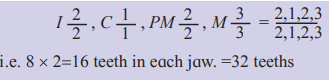
- Teeth: 32 in adults, described as thecodont (fixed in jaw sockets by gomphosis joints), diphyodont (two sets: milk and permanent), and heterodont (four types: incisors, canines, premolars, molars).
- Dental Formula:
- Adult:

- Each jaw has 16 teeth (8 incisors, 4 canines, 8 premolars, 12 molars).
- Tongue: A muscular, triangular organ with papillae (some bearing taste buds) on its upper surface. It aids in food manipulation and swallowing.
- Tooth Anatomy:
- Crown: Visible part above the gum, covered by enamel (hardest substance, made of calcium phosphate and carbonate).
- Root: Embedded in the gum, covered by cementum (bone-like, anchors tooth to socket).
- Neck: Junction between crown and root.
- Dentin: Calcified connective tissue forming the tooth’s bulk, enclosing the pulp cavity (contains nerves, blood vessels).
- Root Canal: Extension of the pulp cavity into the root.
- Pharynx:
- A common passage for food and air, divided into:
- Trachea (upper region, opens via glottis, guarded by epiglottis to prevent food entry during swallowing).
- Oropharynx (lower region, opens into the esophagus via the gullet).
- Deglutition (Swallowing): Controlled by both voluntary and involuntary mechanisms.
- A common passage for food and air, divided into:
- Esophagus:
- A 25 cm long, thin, muscular tube behind the trachea, passing through the neck, rib cage, and diaphragm to join the stomach.
- Histology: Lined by mucus cells; has longitudinal and circular muscles.
- Peristalsis: Rhythmic muscle contractions propel food to the stomach.
- Stomach:
- A muscular, J-shaped sac (25-30 cm) in the upper left abdominal cavity, divided into:
- Cardia: Where the esophagus opens, surrounded by the cardiac sphincter (prevents backflow).
- Fundus: Dome-shaped region above and left of cardia.
- Body: Central portion for food storage.
- Pylorus: Narrow region opening into the duodenum, guarded by the pyloric sphincter.
- Functions: Stores food temporarily, churns it, mixes it with gastric juice, and provides a feeling of satiety.
- A muscular, J-shaped sac (25-30 cm) in the upper left abdominal cavity, divided into:
- Small Intestine:
- A 6-meter-long, 2.5 cm broad, coiled tube divided into:
- Duodenum: U-shaped, 26 cm long, below the stomach.
- Jejunum: 2.5 meters long, narrower, coiled middle portion.
- Ileum: 3.5 meters long, broader, highly coiled, opens into the caecum at the ileocaecal junction.
- Support: Held by mesenteries (carry blood vessels, lymph vessels, nerves).
- A 6-meter-long, 2.5 cm broad, coiled tube divided into:
- Large Intestine:
- 1.5 meters long, wider than the small intestine, includes:
- Caecum: A 6 cm blind sac hosting symbiotic microorganisms. The vermiform appendix (vestigial in humans) arises from it.
- Colon: Tube-like, with ascending, transverse, and descending parts, lined by mucosal cells.
- Rectum: Stores faeces temporarily.
- Anus: Terminal opening, guarded by a sphincter, expels faeces via egestion/defaecation.
- 1.5 meters long, wider than the small intestine, includes:
14.2 Histological Structure of Alimentary Canal
The alimentary canal is lined by four layers (from inside to outside):
- Mucosa:
- Innermost layer with goblet cells secreting mucus to lubricate the canal.
- Modifications:
- Stomach: Forms rugae (folds) and gastric glands (secrete gastric juice).
- Small Intestine: Forms villi (finger-like projections with microvilli) and crypts of Lieberkuhn (intestinal glands).
- Villi: Supplied with capillaries and lacteals (lymph vessels) for nutrient absorption.
- Submucosa:
- Loose connective tissue with blood vessels, lymph vessels, and nerves.
- In the duodenum, contains glands (e.g., Brunner’s glands).
- Muscularis:
- Smooth muscles in three layers: longitudinal (outer), circular (middle), oblique (inner, absent in intestine).
- Thicker in the stomach for churning, thinner in the intestine.
- Serosa:
- Outermost layer of squamous epithelium (mesothelium) and connective tissue.
14.3 Digestive Glands
The digestive glands associated with the alimentary canal are:
- Salivary Glands:
- Types and Position:
- Parotid: In front of the ear.
- Submandibular: Below the lower jaw.
- Sublingual: Below the tongue.
- Structure: Contain serous cells (secrete salivary amylase) and mucous cells (secrete mucus).
- Functions: Saliva lubricates food, initiates starch digestion, and contains ** lysozyme** (antibacterial).
- Types and Position:
- Liver:
- Position: Dark reddish-brown gland below the diaphragm, right upper abdominal cavity (1.2-1.5 kg).
- Structure: Bilobed, covered by Glisson’s capsule, divided into hepatic lobules (polygonal units with a central vein, sinusoids, and Kupffer cells for phagocytosis).
- Functions:
- Secretes bile juice (emulsifies fats, makes food alkaline).
- Stores glycogen, detoxifies substances, synthesizes vitamins (A, D, K, B12), produces blood proteins (prothrombin, fibrinogen).
- Bile is stored in the gall bladder and released via the common bile duct.
- Pancreas:
- Position: Leaf-shaped, heterocrine gland in the gap formed by the duodenum’s bend.
- Exocrine Part: Acinar cells secrete pancreatic juice (contains amylase, lipase, trypsinogen, etc.) via the pancreatic duct.
- Endocrine Part: Islets of Langerhans with:
- α-cells: Secrete glucagon.
- β-cells: Secrete insulin.
- δ-cells: Secrete somatostatin.
- Functions: Pancreatic juice digests carbohydrates, proteins, fats, and nucleic acids; hormones regulate blood sugar.
14.4 Physiology of Digestion
Digestion involves mechanical (chewing, churning, peristalsis) and chemical (enzymatic breakdown) processes.
1. Digestion in the Buccal Cavity:
- Mechanical: Teeth crush food; tongue manipulates it; saliva moistens it, forming a bolus.
- Chemical: Saliva (98% water, 2% electrolytes, amylase, lysozyme) initiates digestion:
- Salivary Amylase (Ptyalin): Converts ~30% starch to maltose at pH 6.8.
- Lysozyme: Antibacterial agent.
- Swallowing: Bolus is pushed to the pharynx and esophagus by peristalsis.
2. Digestion in the Stomach:
- Mechanical: Churning by thick muscular walls mixes food with gastric juice.
- Chemical: Gastric glands secrete:
- Mucus: Protects stomach lining.
- HCl: Activates pepsinogen, kills germs, stops salivary amylase.
- Pepsinogen: Converts to pepsin (active at pH 1.8), digests proteins to peptones and proteoses.
- Rennin (in infants): Curdles milk proteins (casein) for pepsin digestion.
- Output: Food becomes chyme (semifluid, acidic mass).
3. Digestion in the Small Intestine:
- Mechanical: Peristalsis mixes chyme with bile juice, pancreatic juice, and intestinal juice.
- Bile Juice (Liver):
- Contains bile pigments (bilirubin, biliverdin), bile salts, cholesterol, phospholipids.
- Functions: Neutralizes chyme acidity, emulsifies fats, activates lipases.
- Pancreatic Juice (Pancreas):
- Contains:
- Pancreatic Amylase: Converts starch/glycogen to disaccharides.
- Lipases: Hydrolyze fats to fatty acids and monoglycerides.
- Trypsinogen: Activated to trypsin by enterokinase (intestinal enzyme), converts proteins/proteoses/peptones to polypeptides.
- Chymotrypsinogen: Activated to chymotrypsin, converts polypeptides to dipeptides.
- Nucleases: Digest nucleic acids to nucleotides.
- Contains:
- Intestinal Juice (Succus Entericus):
- Contains:
- Maltase: Maltose → Glucose.
- Sucrase: Sucrose → Glucose + Fructose.
- Lactase: Lactose → Glucose + Galactose.
- Dipeptidases: Dipeptides → Amino acids.
- Lipases: Emulsified fats → Fatty acids + Monoglycerides.
- Contains:
- Output: Food becomes chyle (alkaline slurry of digested nutrients).
4. Digestion in the Large Intestine:
- Bacterial Action: Ferments undigested carbohydrates/proteins, producing gases (H₂, CO₂, CH₄) and substances like indole, skatole, H₂S (cause faecal odor).
- Vitamins: Bacteria synthesize B vitamins and vitamin K.
- No enzymes are secreted by the large intestine mucosa.
Regulation of Digestion:
- Neural Control: Sight/smell of food triggers saliva; vagus nerve stimulates gastric juice.
- Hormonal Control:
- Gastrin: Stimulates gastric juice.
- Secretin: Inhibits gastric juice, stimulates bile, pancreatic, and intestinal juice.
- Cholecystokinin (CCK): Similar to secretin, induces satiety.
- Gastric Inhibiting Peptide (GIP): Inhibits gastric secretion.
14.5 Absorption, Assimilation, and Egestion
- Absorption:
- Passage of digested nutrients through the alimentary canal’s mucosa into blood/lymph.
- Mechanisms:
- Simple Diffusion: Glucose, amino acids, chloride ions (based on concentration gradient).
- Facilitated Transport: Fructose, some amino acids (via carrier ions like Na⁺).
- Active Transport: Minerals like sodium (requires energy).
- Sites:
- Mouth: Some drugs (e.g., painkillers).
- Stomach: Water, electrolytes, alcohol, aspirin.
- Small Intestine (90% absorption): Glucose, fructose, amino acids, minerals, water-soluble vitamins (in blood capillaries); lipids, fat-soluble vitamins (A, D, E, K) (in lacteals via chylomicrons).
- Large Intestine: Water, electrolytes, some vitamins, drugs.
- Micelles and Chylomicrons: Bile salts form micelles to transport fatty acids/monoglycerides into villi, where they reform into chylomicrons for lymph transport.
- Assimilation:
- Absorbed nutrients become part of protoplasm in tissues.
- Egestion:
- Undigested waste forms faeces (water, salts, mucosal cells, bacteria, undigested food) in the colon.
- Stored in the rectum, expelled via the anus through defaecation (voluntary, controlled by sphincter muscles).
14.6 Nutritional Disorders and Disorders of Digestive System
- Nutritional Disorders:
- Protein Energy Malnutrition (PEM):
- Kwashiorkor (Children 1-3 years):
- Cause: Protein deficiency.
- Symptoms: Underweight, stunted growth, poor brain development, loss of appetite, anaemia, protruding belly, slender legs, bulging eyes, oedema, skin/hair color changes.
- Marasmus (Infants <1 year):
- Cause: Protein and calorie deficiency.
- Symptoms: Loss of subcutaneous fat, prominent ribs, thin limbs, dry/wrinkled skin, weight loss, stopped digestion/absorption, no oedema.
- Causes: Poverty, large families, early weaning, overdiluted milk.
- Prevention: Proper diet, addressing malnutrition-related infections.
- Kwashiorkor (Children 1-3 years):
- Protein Energy Malnutrition (PEM):
- Digestive System Disorders:
- Indigestion:
- Causes: Overeating, inadequate enzyme secretion, spicy food, anxiety, food poisoning.
- Symptoms: Loss of appetite, acidity, heartburn, regurgitation, dyspepsia, stomach pain.
- Prevention: Avoid large/spicy meals, lying down post-meal, smoking, alcohol.
- Constipation:
- Definition: Defaecation less than once per week.
- Symptoms: Abdominal pain, distortion, rarely perforation.
- Causes: Low fiber diet, inadequate fluid intake, inactivity, neurological dysfunction.
- Prevention: Roughage, fluids, exercise.
- Diarrhoea:
- Definition: Loose, watery stools >3 times/day.
- Symptoms: Dehydration, blood in stool, nausea, bloating, fever.
- Causes: Infections, ulcers, colitis, irritable bowel syndrome.
- Jaundice:
- Symptoms: Yellowing of skin/conjunctiva, whitish stool.
- Causes: Excessive red blood cell breakdown, high bilirubin levels, bile flow obstruction.
- Treatment: Supportive care, rest.
- Vomiting:
- Definition: Expulsion of stomach contents due to reverse peristalsis.
- Control: Medulla’s vomiting center, associated with nausea.
- Alcohol-Related Liver Disorders:
- Steatosis (Fatty Liver): Fat accumulation in liver cells.
- Alcoholic Hepatitis: Inflammation due to alcohol.
- Fibrosis and Cirrhosis: Scarring and severe liver damage.
- Indigestion:
Leave a Reply