
Plant Tissues and Anatomy
8.1 Tissue
Plant tissues are classified based on their ability to divide:
- Meristematic Tissue: Actively dividing cells responsible for growth.
- Permanent Tissue: Mature cells that have lost the ability to divide and perform specialized functions.
8.2 Meristematic Tissue
Meristematic tissues consist of young, actively dividing cells located in specific regions of the plant. These cells are immature, with high metabolic rates and the ability to divide.
Characteristics of Meristematic Tissue:
- Composed of young, living cells.
- Polyhedral or isodiametric in shape, with no intercellular spaces.
- Thin, elastic cell walls made primarily of cellulose.
- Dense protoplasm with a prominent central nucleus.
- Small or absent vacuoles.
- High metabolic activity due to rapid cell division.
Classification of Meristematic Tissue:
- Based on Origin:
- Primordial Meristem (Promeristem): Embryonic meristem occupying a small area at the tips of roots and shoots; also called embryonic meristem.
- Primary Meristem: Originates from primordial meristem; found at root and shoot apices; produces permanent tissues like epidermis, cortex, and vascular tissues.
- Secondary Meristem: Develops from living permanent tissues later in plant growth; always lateral in position (e.g., fascicular cambium, interfascicular cambium, cork cambium).
- Based on Position:
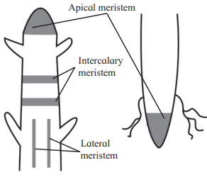
- Apical Meristem: Located at the apices of roots, shoots, and lateral branches; responsible for increasing plant length (called apical initials). Shoot apical meristem is terminal; root apical meristem is subterminal (behind the root cap).
- Intercalary Meristem: Found at the base or top of nodes, primarily in monocots; involved in internode elongation; short-lived.
- Lateral Meristem: Positioned along the sides of the central axis; increases girth (e.g., intrafascicular cambium in vascular bundles of dicots and gymnosperms).
- Based on Function:
- Protoderm: Forms the protective epidermis around organs.
- Procambium: Develops primary vascular tissues (xylem and phloem).
- Ground Meristem: Forms ground tissues like cortex, endodermis, pericycle, pith, and medullary rays.
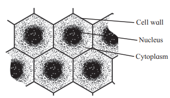
Diagram: Meristematic Tissue
Meristematic Cells: Show thin cell walls, dense cytoplasm, and a central nucleus (Fig. 8.1).
Location of Meristematic Tissue: Apical, intercalary, and lateral meristems (Fig. 8.2).
Can You Tell?
- Characteristics of Meristematic Tissue: Listed above.
- Classification Based on Origin: Primordial, primary, and secondary meristems.
8.3 Permanent Tissue
Permanent tissues are mature cells that have lost the ability to divide, acquiring permanent size, shape, and functions due to morphological, physiological, and functional changes during maturation. They are classified into simple and complex permanent tissues.
A. Simple Permanent Tissues
Composed of one type of cell performing similar functions; can be living or dead.
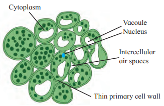
Parenchyma:
- Structure: Thin-walled, isodiametric, round, oval, polygonal, or elongated cells; cell walls made of cellulose; living cells with prominent nucleus, dense cytoplasm, and large vacuoles.
- Intercellular Spaces: Present, but cells may be compact; connected via plasmodesmata, forming a continuous tissue.
- Distribution: Found in epidermis, cortex, pericycle, pith, mesophyll cells, endosperm, xylem, and phloem.
- Functions:
- Stores food (e.g., starch) and water.
- Facilitates gaseous exchange and buoyancy.
- Performs photosynthesis in chlorenchyma (parenchyma with chloroplasts).
- Dedifferentiates to form vascular cambium and cork cambium during secondary growth.
- Diagram: Shows thin cell walls, large vacuoles, and intercellular spaces (Fig. 8.3).
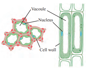
Collenchyma:
- Structure: Living cells with unevenly thickened cell walls (cellulose and pectin, especially at corners); may have pits; cells are circular, oval, or angular in transverse section; small vacuoles; no intercellular spaces, appearing compact.
- Distribution: Found in young stems and petioles; absent in monocots and dicot roots.
- Functions:
- Provides mechanical strength to young stems and petioles.
- Allows bending and pulling without tearing (e.g., in leaves).
- Supports growth and elongation of organs.
- Diagram: Shows unevenly thickened cell walls (Fig. 8.4).
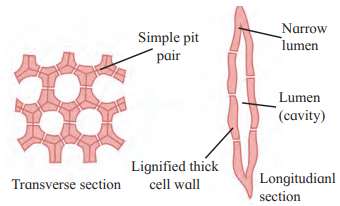
Sclerenchyma:
- Structure: Dead cells with thick, lignified walls; living at production but dead at maturity due to lignin deposition; connected via pits. Two types:
- Fibres: Elongated, thread-like, with tapering, interlocking end walls; narrow, oblique, unbranched pits; arranged in bundles.
- Sclereids: Broad, with blunt end walls; deep, branched, straight pits; occur singly or in loose groups; formed by secondary thickening of parenchyma.
- Distribution: Found in veins, hard seeds, fruit coats, and vascular tissues.
- Functions:
- Provides mechanical strength and rigidity to leaves, epicarps, and seeds.
- Fibres used commercially for products like jute, flax, and hemp.
- Diagram: Shows thick, lignified walls (Fig. 8.5).
B. Complex Permanent Tissues
Composed of multiple cell types functioning as a unit; involved in conduction.
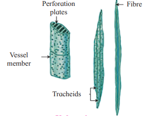
Xylem:
- Components: Tracheids, vessels, xylem parenchyma, xylem fibres.
- Functions: Conducts water and minerals (hadrome); provides mechanical strength.
- Details:
- Tracheids: Elongated, dead, tubular cells with thick, lignified walls; oblique, tapering ends; contribute 95% of wood in gymnosperms, 5% in angiosperms; thickening patterns include:
- Annular: Ring-like.
- Spiral: Helix-like.
- Scalariform: Ladder-like.
- Pitted: Small circular areas (simple or bordered; most advanced).
- Vessels: Formed by end-wall dissolution of vessel elements; wider lumen than tracheids; angular in dicots, rounded in monocots; similar thickening patterns.
- Xylem Parenchyma: Living cells; store food (starch, tannins); facilitate lateral/radial conduction.
- Xylem Fibres: Sclerenchymatous; elongated, spindle-shaped, with lignified walls; provide mechanical support (wood fibres).
- Tracheids: Elongated, dead, tubular cells with thick, lignified walls; oblique, tapering ends; contribute 95% of wood in gymnosperms, 5% in angiosperms; thickening patterns include:
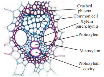
- Types:
- Protoxylem: First-formed, smaller vessels with annular/spiral thickenings.
- Metaxylem: Later-formed, larger vessels with reticulate/pitted thickenings.
- Arrangement:
- Endarch: Protoxylem towards pith, metaxylem towards periphery (stems).
- Exarch: Protoxylem towards periphery, metaxylem towards pith (roots).
- Diagram: Shows xylem components and vascular bundle (Fig. 8.6); tracheid thickenings (Fig. 8.7).
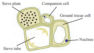
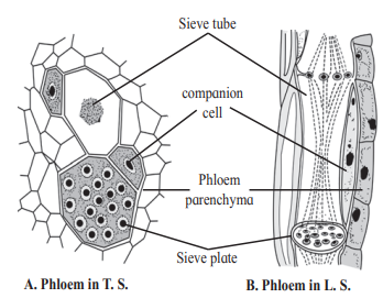
Phloem:
- Components: Sieve tubes, sieve cells, companion cells, phloem parenchyma, phloem fibres.
- Functions: Conducts organic food from source (leaves) to sink (other parts); called leptome.
- Details:
- Sieve Tubes: Long, tubular, living cells with sieve plates (porous septa); lack nucleus at maturity; connected to companion cells via plasmodesmata; found in angiosperms.
- Sieve Cells: Found in lower plants (pteridophytes, gymnosperms); narrow, elongated, with lateral sieve areas.
- Companion Cells: Narrow, living cells with dense cytoplasm and prominent nucleus; regulate sieve tube functions; derived from the same cell as sieve tubes, so death of one causes death of the other.
- Phloem Parenchyma: Living, elongated cells; store food, latex, resins, mucilage; facilitate lateral conduction; absent in most monocots.
- Phloem Fibres: Dead, sclerenchymatous; provide mechanical support; found in secondary phloem; used in ropes and rough clothes.
- Types: Protosieve (first-formed) and metasieve (later-formed).
- Diagram: Shows phloem in transverse and longitudinal sections (Fig. 8.8, 8.9).
Can You Tell?
Parenchyma: Described above.
Sclerenchyma Fibres: Thread-like, elongated, with lignified walls; narrow, oblique pits; provide mechanical strength; used commercially.
T.S. of Phloem Tissue: Shows sieve tubes, companion cells, phloem parenchyma, and fibres (Fig. 8.9).
8.4 Tissue Systems
Plant tissues are organized into three systems based on structure and location:
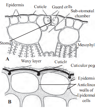
Epidermal Tissue System:
- Components:
- Epidermis: Outermost layer of compact, living cells with a central vacuole, thin cytoplasm, and nucleus; covered by a waxy cuticle to prevent water loss.
- Root Epidermis: Bears unicellular root hairs for absorption.
- Stem Epidermis: Bears multicellular trichomes (branched or unbranched, stiff or soft, secretory) to reduce transpiration.
- Stomata: Pores guarded by specialized guard cells (kidney-shaped in dicots, dumbbell-shaped in monocots); surrounded by subsidiary cells, forming the stomatal apparatus.
- Guard cells contain chloroplasts and regulate opening/closing via turgor pressure.
- Facilitate gas exchange and water vapor loss.
- Epidermis: Outermost layer of compact, living cells with a central vacuole, thin cytoplasm, and nucleus; covered by a waxy cuticle to prevent water loss.
- Diagram: Shows epidermal cells, stomata, and cuticle (Fig. 8.10, 8.11).

Ground Tissue System:
- Components: All tissues except epidermal and vascular tissues; includes:
- Parenchyma in cortex, pericycle, pith, and medullary rays.
- Collenchyma in hypodermis.
- Sclerenchyma in hypodermis.
- Chloroplast-containing mesophyll in leaves.
- Functions: Storage, photosynthesis, mechanical support.
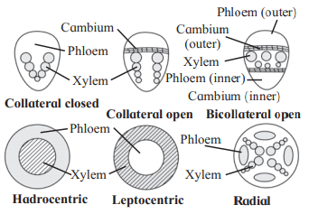
Vascular Tissue System:
- Components: Xylem and phloem arranged in vascular bundles.
- Types:
- Radial: Xylem and phloem on separate radii (common in roots).
- Conjoint, Collateral: Xylem and phloem on the same radius, with xylem inside and phloem outside (stems).
- Open: Cambium present between xylem and phloem (dicots; allows secondary growth).
- Closed: Cambium absent (monocots; no secondary growth).
- Bicollateral: Phloem on both sides of xylem with intervening cambium (e.g., Cucurbitaceae).
- Concentric:
- Hadrocentric: Xylem surrounds phloem.
- Leptocentric: Phloem surrounds xylem.
- Amphicribral: Xylem encircled by phloem on both faces.
- Amphivasal: Phloem encircled by xylem on both faces.
- Diagram: Shows vascular bundle types (Fig. 8.12).
8.5 Secondary Growth in Plants
Secondary growth increases the girth of stems and roots in dicots and gymnosperms, driven by lateral meristems (vascular cambium and cork cambium).
Secondary Growth in Stems:
- Formation of Cambial Ring:
- Intrafascicular Cambium: Present between primary xylem and phloem in vascular bundles; becomes active in favorable seasons.
- Interfascicular Cambium: Formed when ray parenchyma cells (fusiform and ray initials) dedifferentiate and become meristematic.
- These join to form a continuous cambial ring in one plane.
- Activity: The cambial ring cuts off new cells:
- Inner Side: Matures into secondary xylem (more abundant).
- Outer Side: Matures into secondary phloem (less abundant).
- Outcome: Increases stem girth, supports conduction of water, minerals, and food, and provides mechanical strength.
- Diagram: Shows secondary growth in dicot stem (Fig. 8.13).

Secondary Growth in Roots:
- Process: Conjunctive parenchyma cells near primary phloem bundles become meristematic, forming vascular cambium. This produces:
- Secondary Xylem: On the inner side.
- Secondary Phloem: On the outer side.
- Periderm Formation: Cork cambium forms in the pericycle, producing phellem and phelloderm.
- Outcome: Similar to stem, increases root girth and supports conduction.
8.6 Wood
Wood is secondary xylem formed during secondary growth, with seasonal variations:
- Spring Wood (Early Wood):
- Formed in favorable conditions (spring).
- Broader xylem bands, lighter color, thin-walled tracheids, wide lumen, fewer fibres, low density.
- Autumn Wood (Late Wood):
- Formed in unfavorable conditions (autumn).
- Narrow xylem bands, darker color, thick-walled tracheids, narrow lumen, abundant fibres, high density.
- Heartwood (Duramen):
- Inner, non-functional, durable wood.
- Tracheary elements plugged by tyloses (parenchyma in-growths).
- Filled with extractives (oils, gums, resins, tannins), making it resistant to pathogens.
- Sapwood (Alburnum):
- Outer, functional wood with living cells.
- No deposition of extractives; lighter, less durable, more susceptible to pathogens.
- Conducts sap (water and minerals).
8.7 Cork Cambium and Secondary Growth
- Cork Cambium (Phellogen):
- Develops in the outer cortical layer when the epidermis ruptures due to secondary growth.
- Outer cortical cells become meristematic, producing:
- Phellem (Cork): On the outer side; impervious due to suberized walls.
- Phelloderm (Secondary Cortex): On the inner side; parenchymatous.
- Phellogen, phellem, and phelloderm constitute the periderm.
- Bark: Non-technical term for all tissues external to vascular cambium, including secondary phloem.
- Early-Season Bark: Soft.
- Late-Season Bark: Hard.
- Lenticels: Aerating pores on the bark surface (raised scars); formed by increased phellogen activity; facilitate gas and water vapor exchange.
- Anomalous Secondary Growth: Occurs in some monocots (e.g., Dracaena, Agave, palms, sweet potato roots) due to accessory cambium, despite lacking typical cambium.
8.8 Anatomy of Root, Stem, and Leaf
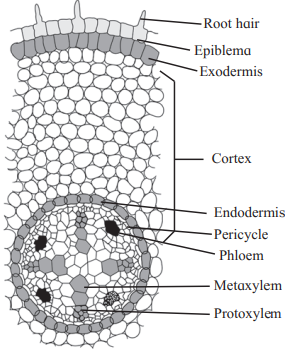
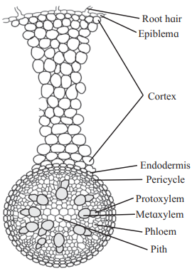
A. Anatomy of Dicot Root
- Epiblema: Outermost single layer without cuticle; some cells form unicellular root hairs for absorption.
- Cortex: Several layers of parenchymatous cells; stores food and water.
- Exodermis: Outer cortical layer becomes cutinized after epiblema dies.
- Endodermis: Innermost cortical layer with barrel-shaped cells; radial walls bear Casparian strips (suberin bands); unthickened passage cells near protoxylem.
- Pericycle: Single layer of parenchyma below endodermis; bounds the stele.
- Stele (Vascular Cylinder):
- Contains 2-6 radial vascular bundles (diarch to hexarch).
- Xylem is exarch (protoxylem towards periphery).
- Parenchymatous conjunctive tissue between xylem and phloem.
- Pith: Narrow, central parenchymatous region with or without intercellular spaces.
- Secondary Growth: Cambium ring forms between xylem and phloem, producing secondary tissues.
- Diagram: T.S. of dicot root (Fig. 8.14).
B. Anatomy of Monocot Root
- Similar to dicot root but with:
- Polyarch Condition: More than 6 xylem bundles.
- Large, Well-Developed Pith.
- No Secondary Growth due to absence of cambium.
- Diagram: T.S. of monocot root (Fig. 8.15).
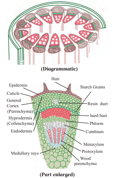
C. Anatomy of Dicot Stem (e.g., Sunflower)
- Epidermis: Single layer with multicellular trichomes and a cuticle.
- Cortex:
- Hypodermis: 3-5 layers of collenchyma; no intercellular spaces.
- General Cortex: Several layers of large parenchymatous cells with intercellular spaces.
- Endodermis: Innermost layer of barrel-shaped cells; also called starch sheath.
- Stele:
- Pericycle: Multilayered sclerenchyma (hard bast) near vascular bundles.
- Vascular Bundles: Conjoint, collateral, open; arranged in a ring; xylem is endarch; cambium present between xylem and phloem.
- Pith: Central, large parenchymatous region with intercellular spaces.
- Diagram: T.S. of dicot stem (Fig. 8.16).
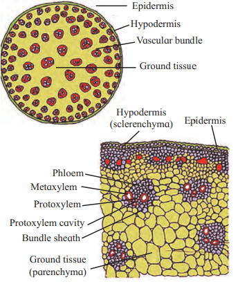
D. Anatomy of Monocot Stem
- Epidermis: Single layer without trichomes.
- Hypodermis: Sclerenchymatous.
- Ground Tissue: Parenchymatous; contains numerous scattered vascular bundles.
- Vascular Bundles: Conjoint, collateral, closed (no cambium); surrounded by sclerenchymatous bundle sheath; xylem is endarch with a lysigenous cavity.
- Pith: Absent.
- Secondary Growth: Absent.
- Diagram: T.S. of monocot stem (Fig. 8.17).
E. Anatomy of Dicot Leaf (Dorsiventral)
- Upper Epidermis: Single layer of tightly packed, rectangular, chloroplast-free cells; thick cuticle; stomata usually absent.
- Mesophyll:
- Palisade Parenchyma: Below upper epidermis; closely packed, elongated cells with abundant chloroplasts; primary site of photosynthesis.
- Spongy Parenchyma: Below palisade; loosely arranged, irregular cells with intercellular spaces and chloroplasts; connected to stomata for gas exchange.
- Vascular System: Closed vascular bundles (no cambium); xylem towards upper epidermis, phloem towards lower; surrounded by parenchymatous bundle sheath.
- Lower Epidermis: Single layer of compact cells; thin cuticle; contains numerous stomata with sub-stomatal chambers.
- Diagram: T.S. of dicot leaf (Fig. 8.17).
F. Anatomy of Monocot Leaf (Isobilateral)
- Similar to dicot leaf but:
- Stomata: Present on both surfaces (amphistomatic).
- Mesophyll: Not differentiated into palisade and spongy tissue; uniform parenchyma with chloroplasts.
- Veins: Parallel.
- Bulliform Cells: Present; help in leaf rolling to reduce water loss.
- Diagram: T.S. of monocot leaf (Fig. 8.18).
Leave a Reply