
Skeleton and Movement
1. Choose the correct option
A. The functional unit of striated muscle is …………..
a. cross bridges b. myofibril
c. sarcomere d. z-band
Answer: c. sarcomere
(The sarcomere is the contractile unit of striated muscle, containing actin and myosin filaments responsible for muscle contraction.)
B. A person slips from the staircase and breaks his ankle bone. Which bones are involved?
a. Carpals b. Tarsal
c. Metacarpals d. Metatarsals
Answer: b. Tarsal
(The ankle consists of tarsal bones, which can be fractured in such an injury.)
C. Muscle fatigue is due to accumulation of ……..
a. pyruvic acid b. lactic acid
c. malic acid d. succinic acid
Answer: b. lactic acid
(Lactic acid accumulates during anaerobic glycolysis, causing muscle fatigue.)
D. Which one of the following is NOT antagonistic muscle pair?
a. Flexo-extensor
b. Adductor-abductor
c. Levator-depressor
d. Sphinetro-suprinater
Answer: d. Sphinctro-suprinater
(Sphincters and supinators do not form an antagonistic pair, unlike flexor-extensor, adductor-abductor, or levator-depressor.)
E. Swelling of sprained foot is reduced by soaking in hot water containing a large amount of common salt,
a. due to osmosis
b. due to plasmolysis
c. due to electrolysis
d. due to photolysis
Answer: a. due to osmosis
(The high salt concentration draws water out of the swollen tissue via osmosis, reducing swelling.)
F. Role of calcium in muscle contraction is ……….
a. to break the cross bridges as a cofactor in the hydrolysis of ATP
b. to bind with troponin, changing its shape so that the actin filament is exposed
c. to transmit the action potential across the neuromuscular junction.
d. to re-establish the polarisation of the plasma membrane following an action potential
Answer: b. to bind with troponin, changing its shape so that the actin filament is exposed
(Calcium binds to troponin, causing a conformational change that moves tropomyosin, exposing actin’s binding sites for myosin.)
G. Hyper-secretion of parathormone can cause which of the following disorders?
a. Gout b. Rheumatoid arthritis
c. Osteoporosis d. Gull’s disease
Answer: c. Osteoporosis
(Excess parathormone increases bone resorption, leading to porous and brittle bones, characteristic of osteoporosis.)
H. Select correct option between two nasal bones
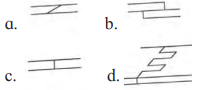
Answer:
![]()
2. Answer the following questions
A. What kind of contraction occurs in your neck muscles while you are reading your class assignment?
Answer: Isometric contraction occurs in the neck muscles. These muscles contract to hold the head in a stable position without causing movement, maintaining posture during reading.
B. Observe the diagram and enlist importance of ‘A’, ‘B’ and ‘C’
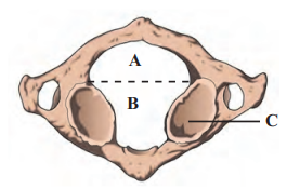
Answer:
A – Posterior portion of vertebral foramen of atlas vertebrae; Importance – The spinal cord runs through this portion of vertebral foramen
B – Anterior portion of vertebral foramen of axis vertebrae; Importance – In this portion, the odontoid process of axis vertebrae forms ‘NO’ joint.
C – Inferior articular facet; Importance – It articulates with superior articular facet of axis and permits rotatory movement of head.
C. Raju intends to train biceps; while exercising using dumbbells, which joints should remain stationary and which should move?
Answer:
- Stationary joint: The shoulder joint should remain relatively stationary to stabilize the upper arm.
- Moving joint: The elbow joint should move, allowing flexion and extension as the biceps contract and relax during dumbbell exercises.
D. In a road accident, Moses fractured his leg. One of the passers by, tied a wodden plank to the fractured leg while Moseswas rushed to the hospital Was this essential? Why?
Answer: Yes, this was essential. Tying a wooden plank acts as a splint, immobilizing the fractured leg to prevent further movement of the broken bones. This reduces pain, prevents additional tissue damage, and minimizes the risk of complications like blood vessel or nerve injury during transport to the hospital.
E. Sprain is more painful than fracture. Why?
Answer: A sprain can be more painful than a fracture because it involves damage to ligaments, which are richly supplied with nerves, leading to intense pain. Fractures may sometimes cause less immediate pain if the break is clean and does not involve significant soft tissue damage. Additionally, sprains often result in swelling and inflammation, which further stimulate pain receptors.
F. Why a red muscle can work for a prolonged period whereas white muscle fibre suffers from fatigue after a shorter work? (Refer to chapter animal tissues.)
Answer: Red muscle fibers (slow-twitch) are rich in myoglobin and mitochondria, enabling efficient aerobic respiration and sustained energy production, allowing them to work for prolonged periods without fatigue. White muscle fibers (fast-twitch) rely on anaerobic glycolysis, leading to rapid energy production but quick accumulation of lactic acid, causing fatigue after shorter durations.
3. Answer the following questions in detail
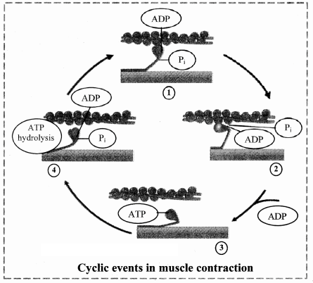
A. How is the structure of sarcomere suitable for the contractility of the muscle? Explain its function according to sliding filament theory. (Refer to chapter animal tissues.)
Answer: The sarcomere, the functional unit of striated muscle, is highly organized to facilitate muscle contraction. Its structure includes:
- Actin filaments (thin filaments) anchored at the Z-lines, forming the boundaries of the sarcomere.
- Myosin filaments (thick filaments) located centrally, with heads that form cross-bridges with actin.
- Tropomyosin and troponin on actin regulate the interaction with myosin.
Sliding Filament Theory:
According to H.E. Huxley and A.F. Huxley’s sliding filament theory, muscle contraction occurs when actin and myosin filaments slide past each other, shortening the sarcomere without changing the length of the filaments themselves. The process involves:
- Action Potential: A nerve impulse triggers the release of calcium ions from the sarcoplasmic reticulum into the sarcoplasm.
- Calcium Binding: Calcium binds to troponin, causing tropomyosin to shift and expose actin’s binding sites.
- Cross-Bridge Formation: Myosin heads, energized by ATP hydrolysis, bind to actin, forming an actomyosin complex.
- Power Stroke: Myosin heads tilt, pulling actin filaments inward, shortening the sarcomere.
Detachment and Reset: ATP binds to myosin, releasing it from actin, and is hydrolyzed to re-energize the myosin head for another cycle. - This cyclic interaction, driven by ATP, results in muscle contraction as sarcomeres shorten across the muscle fiber.
B. Ragini, a 50 year old office goer, suffered hair-line cracks in her right and left foot in short intervals of time. She was worried about minor jerks leading to hair line cracks in bones. Doctor explained to her why it must be happening and prescribed medicines.
What must be the cause of Ragini’s problem? Why has it occurred? What precautions she should have taken earlier? What care she should take in future?
Answer:
- Cause: Ragini’s hairline cracks are likely due to osteoporosis, a condition where bones become porous and brittle, making them prone to fractures from minor stress.
- Reason for Occurrence: At 50, Ragini is likely postmenopausal, leading to decreased estrogen levels, which accelerates bone resorption over formation. Other factors may include inadequate calcium and vitamin D intake, lack of weight-bearing exercise, or genetic predisposition.
- Precautions She Should Have Taken Earlier:
- Consumed a diet rich in calcium (e.g., dairy, leafy greens) and vitamin D (e.g., fish, sunlight exposure).
- Engaged in regular weight-bearing exercises (e.g., walking, strength training) to strengthen bones.
- Avoided smoking and excessive alcohol, which weaken bones.
- Undergone bone density screenings to detect early bone loss.
- Future Care:
- Take prescribed medications (e.g., bisphosphonates) to slow bone loss.
- Follow a calcium- and vitamin D-rich diet.
- Perform low-impact exercises to improve bone strength and balance, reducing fall risk.
- Schedule regular bone density tests to monitor bone health.
C. How does structure of actin and myosin help muscle contraction?
Answer: Actin Filament Structure:
- Composed of F-actin (double-stranded polymer of G-actin molecules, each with an ADP molecule).
- Tropomyosin wraps around F-actin, covering myosin-binding sites in the resting state.
Troponin, a complex of three proteins, binds to tropomyosin and actin, with a high affinity for calcium ions. - This structure allows actin to interact with myosin only when activated by calcium, ensuring controlled contraction.
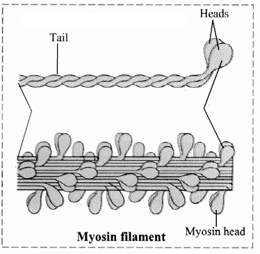
Myosin Filament Structure:
- Composed of multiple meromyosin units, each with two heavy chains (forming a double helix with a tail and head) and four light chains.
- The myosin head has ATPase activity, splitting ATP to provide energy, and forms cross-bridges with actin.
- Myosin tails align toward the sarcomere’s center, with heads projecting outward for actin interaction.
Role in Contraction:
When calcium binds to troponin, it shifts tropomyosin, exposing actin’s binding sites. Myosin heads, energized by ATP hydrolysis, bind to actin, forming cross-bridges. The heads tilt (power stroke), pulling actin filaments inward, shortening the sarcomere. ATP then detaches myosin, allowing the cycle to repeat, driving muscle contraction.
D. Justify the structure of atlas and axis vertebrae with respect to their position and function.
Answer: Atlas Vertebra (1st Cervical):
- Structure: Ring-like, lacking a centrum and spinous process, with large transverse processes and a vertebral foramen divided by a transverse ligament. It has facets for articulation with the occipital condyles of the skull.
- Position and Function: Located at the top of the vertebral column, it supports the skull and forms the “Yes joint” (atlanto-occipital joint), allowing nodding movements (flexion and extension). Its ring-like structure and large foramen accommodate the spinal cord and allow rotational flexibility.
Axis Vertebra (2nd Cervical):
- Structure: Features an odontoid process (a tooth-like projection from the centrum) that fits into the atlas’s vertebral foramen, forming a pivot joint. It has a robust spinous process and transverse foramina.
- Position and Function: Positioned below the atlas, it enables the “No joint” (atlanto-axial joint), allowing side-to-side head rotation. The odontoid process acts as a pivot, while the sturdy structure supports rotational stability.
Together, the atlas and axis facilitate head movements (nodding and rotation) while protecting the spinal cord, with their specialized structures tailored to their roles in the cervical region.
E. Observe the blood report given below and diagnose the possible disorder.
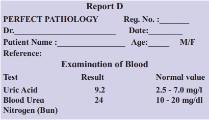
Answer: On observing Report D, it is clear that the level of uric acid is more than normal, thus the patient must be suffering from gouty arthritis.
Also, the elevated blood urea nitrogen (BUN) indicates dysfunctional liver and/ or kidneys. It generally occurs due to decrease in GFR, caused by renal disease or obstruction of urinary tract.
4. Write short notes on following points
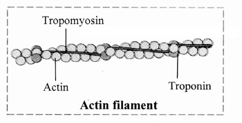
A. Actin filament
Answer: Actin filaments (thin filaments) are contractile proteins in the sarcomere, essential for muscle contraction. They consist of:
- F-actin: A double-stranded polymer of G-actin molecules, each with an ADP molecule, forming the filament’s backbone.
- Tropomyosin: Two protein strands loosely wrapped around F-actin, covering myosin-binding sites in the resting state.
- Troponin: A three-protein complex attached to tropomyosin, with affinity for calcium ions.
When calcium binds to troponin, tropomyosin shifts, exposing actin’s binding sites for myosin, enabling cross-bridge formation and muscle contraction.
B. Myosin filament
Answer: Myosin filaments (thick filaments) are key contractile proteins in the sarcomere. Each filament is composed of multiple meromyosin units, with:
- Heavy chains: Two chains coiled into a double helix, forming a long tail and a globular head with ATPase activity.
- Light chains: Four per myosin molecule, associated with the head, stabilizing its structure.
Cross-bridges: Myosin heads project outward, binding to actin during contraction. - Myosin heads hydrolyze ATP to provide energy, tilt to pull actin filaments (power stroke), and detach with new ATP, driving muscle contraction.
C. Role of calcium ions in contraction and relaxation of muscles.
Answer:
- Contraction: Calcium ions are released from the sarcoplasmic reticulum when a nerve impulse arrives. They bind to troponin, causing a conformational change that shifts tropomyosin, exposing actin’s myosin-binding sites. This allows myosin heads to form cross-bridges with actin, leading to muscle contraction via the sliding filament mechanism.
- Relaxation: When stimulation ceases, calcium ions are actively pumped back into the sarcoplasmic reticulum using ATP. This removes calcium from troponin, restoring the troponin-tropomyosin complex, which covers actin’s binding sites. Myosin detaches from actin (using ATP), and actin filaments slide back, relaxing the muscle. Calcium thus regulates the interaction between actin and myosin, controlling contraction and relaxation.
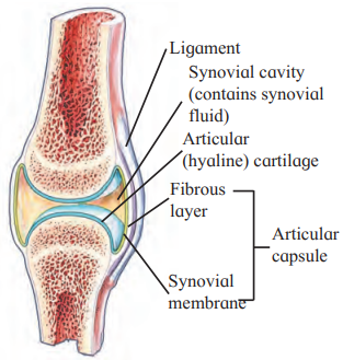
5. Draw labelled diagrams
A. Synovial joint
B. Different cartilagenous joints.
Leave a Reply