
Plant Tissues and Anatomy – Solutions
1. Choose the correct option
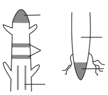
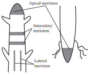
A. Based on location or position of meristematic regions are divided into ………………. types
a. one b. two
c. three d. none of the above
Answer: c. three
- Explanation: Meristematic regions are classified based on location into three types: apical, intercalary, and lateral meristems
B. Cambium is also called …………….
a. apical meristem
b. intercalary meristem
c. lateral meristem
d. none of the above
Answer: c. lateral meristem
- Explanation: Cambium, such as intrafascicular and interfascicular cambium, is classified as a lateral meristem because it is located along the sides of the central axis and contributes to increasing the girth of stems or roots
C. Collenchyma is a type of …………….. tissue.
a. living b. dead
c. living and dead d. none of the above
Answer: a. living
- Explanation: Collenchyma is a simple permanent tissue made up of living cells with unevenly thickened cell walls, providing mechanical support.
D. ………………… is a complex permanent tissue.
a. Parenchyma b. Sclerenchyma
c. Chlorenchyma d. Xylem
Answer: d. Xylem
- Explanation: Xylem is a complex permanent tissue composed of multiple cell types (tracheids, vessels, xylem parenchyma, and xylem fibres) that function together for conduction and support.
E. Mesophyll tissue is present in…………….
a. root b. stem
c. leaf d. flower
Answer: c. leaf
- Explanation: Mesophyll tissue, consisting of palisade and spongy parenchyma, is found in leaves where it facilitates photosynthesis
2. Answer the following questions
A. A fresh section was taken by a student but he was very disappointed because there were only few green and most colourless cells. Teacher provided a pink colour solution. The section was immersed in this solution and when observed it was much clearer. What is the magic?
Answer: The pink colour solution is likely a staining agent, such as safranin, used to enhance the visibility of cell structures under a microscope. Many plant cells, especially non-photosynthetic ones, appear colourless in fresh sections due to the absence of pigments like chlorophyll. The stain binds to cell components (e.g., cell walls, nuclei), making them distinct and easier to observe, thus clarifying the section’s anatomical details.
B. While observing a section many scattered vascular bundles could be seen. Teacher opined that in spite of this large number the stem cannot grow in girth. Why?
Answer: The stem is likely a monocot stem, which has scattered vascular bundles that are conjoint, collateral, and closed (lacking cambium). The absence of cambium prevents secondary growth, so the stem cannot increase in girth.
C. A section of the stem had vascular bundles, where one vascular tissue was wrapped around the other. How will you technically describe it?
Answer: This is a concentric vascular bundle, where one vascular tissue (e.g., xylem or phloem) completely encircles the other. It can be described as hadrocentric (xylem encircled by phloem) or leptocentric (phloem encircled by xylem), or further as amphicribral (xylem encircled by phloem on both faces) or amphivasal (phloem encircled by xylem on both faces).
D. There were two cut logs of wood lying in the campus. One had growth rings and other didn’t. Teacher said it is due to differences in their pattern of growth which is dependent on season. How?
Answer: The log with growth rings is likely from a dicot or gymnosperm, which undergoes secondary growth due to vascular cambium activity. Growth rings form due to seasonal variations, with spring wood (broader, lighter, thin-walled tracheids) and autumn wood (narrower, darker, thick-walled tracheids) produced in favorable and unfavorable seasons, respectively. The log without growth rings is likely from a monocot, which lacks cambium and secondary growth, so no seasonal rings are formed.
E. While on the trip to Kashmir, Pintoo observed that cut portions of large trees show distinct rings, which he never found in Maharashtra. Why is so?
Answer: In Kashmir, a region with distinct seasonal changes (e.g., cold winters and warm summers), trees (likely dicots or gymnosperms) exhibit secondary growth with clear growth rings due to seasonal cambial activity producing spring and autumn wood. In Maharashtra, with a more tropical or less seasonally variable climate, trees may not form distinct rings, or the trees observed (possibly monocots) lack secondary growth, resulting in no rings.
F. A student was observing a slide with no label. The section had some vascular bundles scattered in the ground tissue. It is section of a monocot stem! He exclaimed. No! it is section of fern rachis, said the teacher. Teacher told to observe vascular bundle again. Student agreed, Why?
Answer: The student mistook the scattered vascular bundles for a monocot stem, which typically has such an arrangement. However, fern rachis (the main axis of a fern frond) also has scattered vascular bundles, but their structure differs. Fern vascular bundles may have different arrangements or components (e.g., lacking vessels, having tracheids typical of pteridophytes) compared to monocot stems, which have closed, conjoint, collateral bundles with vessels. The teacher’s instruction to re-observe likely highlighted these structural differences.
G. Student found a wooden stopper in lab. He was told by an old lab attendant that it is there for many years. He kept thinking how it did not rot?
Answer: The wooden stopper is likely made of heartwood (duramen), the inner, non-functional part of secondary xylem. Heartwood is resistant to rot due to the deposition of extractives (oils, gums, resins, tannins) and tyloses, which plug tracheary elements, making it durable and resistant to pathogens and decay.
H. Student while observing a slide of leaf section observed many stomata on the upper surface. He thought he has placed slide upside down. Teacher confirmed it is rightly placed. Explain.
Answer: The leaf is likely an isobilateral leaf, typical of monocots, where stomata are present on both upper (adaxial) and lower (abaxial) surfaces, unlike dorsiventral dicot leaves, which usually have stomata primarily on the lower surface. The presence of stomata on the upper surface is normal for such leaves, confirming the slide was correctly placed.
3. Write short notes on the following points
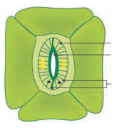
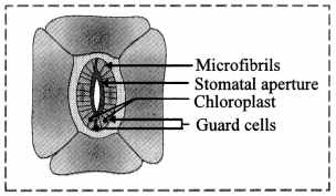
A. Structure of stomata
Answer: Stomata are small gateways in the epidermal cells of leaves and stems, facilitating gas exchange and transpiration. Each stoma is surrounded by two specialized guard cells, which are kidney-shaped in dicots and dumbbell-shaped in monocots. Guard cells contain chloroplasts and regulate stomatal opening and closing by changing turgor pressure. Surrounding the guard cells are subsidiary cells, forming the stomatal apparatus. A sub-stomatal chamber, an air space beneath the stoma, connects to intercellular spaces in the mesophyll, aiding gas exchange.
B. Secondary growth
Answer: Secondary growth refers to the increase in girth of stems and roots in dicotyledonous plants and gymnosperms, driven by lateral meristems (vascular cambium and cork cambium). The vascular cambium, formed from intrafascicular cambium and dedifferentiated medullary ray cells (interfascicular cambium), produces secondary xylem (inward) and secondary phloem (outward). Cork cambium (phellogen) forms phellem (cork) and phelloderm, constituting the periderm. This process replaces the epidermis, adds structural support, and enables conduction, with heartwood and sapwood forming in woody plants.
C. Peculiarity of a sclerenchyma cell wall
Answer: Sclerenchyma cell walls are thick, lignified, and rigid, providing mechanical strength and support. The walls are uniformly thickened due to lignin deposition, making the cells dead at maturity. They feature numerous pits, which are narrow, unbranched, and oblique in fibres, and deep, branched, and straight in sclereids. These pits allow limited intercellular communication. The lignified walls contribute to the tissue’s stiffness and durability, as seen in fibres (e.g., jute, hemp) and sclereids in seeds and epicarps.
4. Differentiate
A. Vascular bundle of monocot and dicot
Answer:
| Feature | Monocot | Dicot |
|---|---|---|
| Arrangement | Scattered in ground tissue. | Arranged in a ring. |
| Type | Conjoint, collateral, closed (no cambium). | Conjoint, collateral, open (cambium present). |
| Bundle Sheath | Surrounded by sclerenchymatous bundle sheath. | Not always surrounded by sclerenchyma, may have parenchymatous sheath. |
| Xylem | Endarch, with lysigenous protoxylem cavity. | Endarch, no lysigenous cavity. |
| Secondary Growth | Absent due to lack of cambium. | Present due to cambium activity. |
B. Xylem and Phloem functioning
Answer:
| Feature | Xylem | Phloem |
|---|---|---|
| Main Function | Conducts water and minerals from roots to other parts. | Conducts organic food (sugars) from leaves (source) to other parts (sink). |
| Components | Tracheids, vessels, xylem parenchyma, xylem fibres. | Sieve tubes, companion cells, phloem parenchyma, phloem fibres |
| Cell Status | Mostly dead (except xylem parenchyma). | Mostly living (except phloem fibres). |
| Direction of Flow | Unidirectional (upward). | Bidirectional (source to sink). |
| Additional Role | Provides mechanical strength. | Stores food, latex, resins, etc., in phloem parenchyma. |
C. Internal or anatomical differences between monocots and dicots leaves
Answer:
| Feature | Monocot Leaf (Isobilateral) | Dicot Leaf (Dorsiventral) |
|---|---|---|
| Mesophyll Differentiation | Not differentiated; uniform parenchymatous cells with chloroplasts. | Differentiated into palisade (upper, tightly packed) and spongy (lower, loosely arranged) parenchyma. |
| Stomata Distribution | Present on both upper and lower epidermis (amphistomatic). | Mostly on lower epidermis, rarely on upper. |
| Guard Cell Shape | Dumbbell-shaped. | Kidney-shaped. |
| Vascular Bundles | Parallel venation, conjoint, collateral, closed. | Reticulate venation, conjoint, collateral, closed. |
| Bulliform Cells | Present, help in leaf rolling. | Absent. |
5. Draw neat labelled diagrams
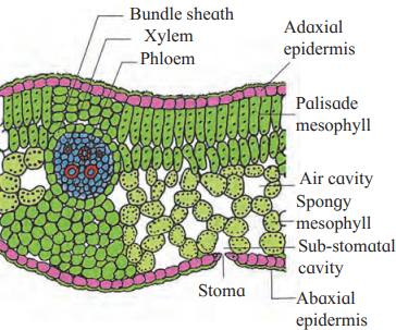
A. T. S. of Dicot leaf.
Answer:
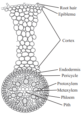
B. T. S. of Monocot root.
Answer:
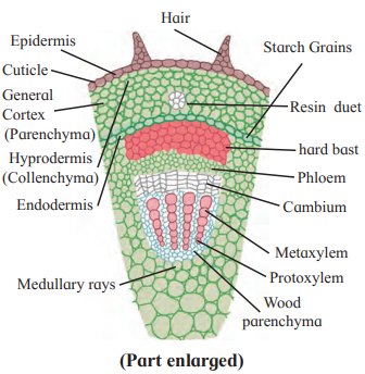
C. T. S. of dicot stem.
Answer:
6. Write the information related parts in the diagrams given below
Answer:
Answer:
7. Identify the following diagrams, label it and prepare a chart of characteristics.
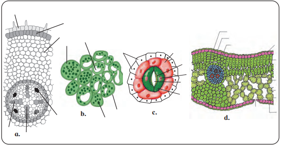
Answer: A.
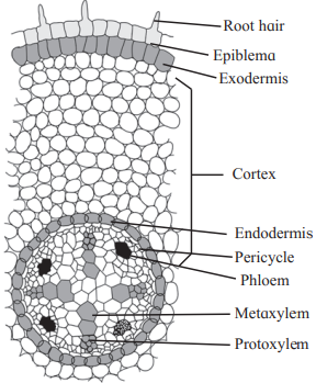
Characteristics of Transverse Section ( T.S.) of Dicot Root-
| Layer/Structure | Characteristics |
|---|---|
| Epiblema |
|
| Cortex |
|
| Exodermis |
|
| Endodermis |
|
| Pericycle |
|
| Stele (Vascular Cylinder) |
|
| Xylem |
|
| Phloem |
|
| Conjunctive Tissue |
|
| Pith |
|
| Cambium |
|
Answer: B.
Characteristics of Simple Permanent Tissue-
| Type of Tissue | Characteristics | Function | Example Locations |
|---|---|---|---|
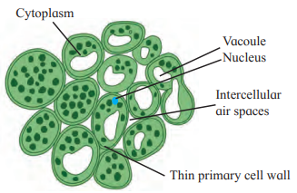
| Parenchyma |
|
|
|
| Collenchyma |
|
|
|
| Sclerenchyma |
|
|
Answer: C.

Chart of Characteristics: Epidermal Cells –
| Feature | Characteristics |
|---|---|
| Origin | Derived from protoderm or dermatogen. |
| Structure |
|
| Cell Wall |
|
| Intercellular Spaces | Absent, cells are tightly packed. |
| Living/Dead | Living cells. |
| Specialized Structures |
|
| Guard Cells |
|
| Location | Forms the outermost covering of the plant body (roots, stems, leaves). |
| Functions |
|
| Variations |
|
Answer: D.
Chart of Characteristics: Transverse Section (T.S.) of Dicot Leaf
| Feature | Characteristics |
|---|---|
| Leaf Type | Dorsiventral, with distinct upper (adaxial) and lower (abaxial) surfaces; upper surface darker (faces sun), lower surface lighter. |
| Upper Epidermis |
|
| Lower Epidermis |
|
| Stomata |
|
| Mesophyll |
|
| Vascular System |
|
| Secondary Growth | Absent due to lack of cambium in vascular bundles. |
| Functions |
|
| Orientation | Typically horizontal, with upper surface facing the sun. |
8. Distinguish between Dicot and Monocot leaf on the basis of following characters.
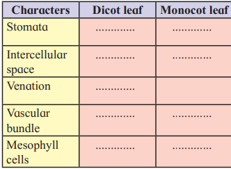
Answer:
| Characters | Dicot leaf | Monocot leaf |
| Stomata | Stomata are restricted to lower epidermis. Guard cells of stoma are kidney shaped. | Stomata occur on both epidermis. Guard cells of stoma are dumbbell shaped. |
| Intercellular space | More intercellular spaces due to presence of spongy parenchyma. | Less intercellular spaces as mesophyll is not differentiated into spongy and palisade tissue. |
| Venation | Reticulate venation | Parallel venation |
| Vascular bundle | Vascular bundles of varying size. The size of the vascular bundles is dependent on the size of the veins which vary in thickness in dicot leaf. | Vascular bundles are nearly of similar size (Except in main veins). |
| Mesophyll cells | Mesophyll tissue is differentiated into palisade parenchyma and spongy parenchyma. | Mesophyll tissue is not differentiated into palisade parenchyma and spongy parenchyma. |
Leave a Reply